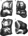
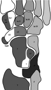
First evidence of a bipartite medial cuneiform in the hominin fossil record: a case report from the Early Pleistocene site of Dmanisi
- PMID: 20579174
- PMCID: PMC2952383
- DOI: 10.1111/j.1469-7580.2010.01236.x
First evidence of a bipartite medial cuneiform in the hominin fossil record: a case report from the Early Pleistocene site of Dmanisi
Abstract
A medial cuneiform exhibiting complete bipartition was discovered at the Early Pleistocene site of Dmanisi, Georgia. The specimen is the oldest known instance of this anatomical variant in the hominin fossil record. Here we compare developmental variation of the medial cuneiform in fossil hominins, extant humans and great apes, and discuss potential implications of bipartition for hominin foot phylogeny and function. Complete bipartition is rare among modern humans (< 1%); incomplete bipartition was found in 2 of 200 examined great ape specimens and also appears in the form of a divided distal articular surface in the Stw573c Australopithecus africanus specimen. Although various developmental pathways lead to medial cuneiform bipartition, it appears that the bipartite bone does not deviate significantly from normal overall morphology. Together, these data indicate that bipartition represents a phyletically old developmental variant of the medial cuneiform, which does not, however, affect the species-specific morphology and function of this bone.
Figures


Similar articles
-
Mandibular size and shape variation in the hominins at Dmanisi, Republic of Georgia.J Hum Evol. 2006 Jul;51(1):36-49. doi: 10.1016/j.jhevol.2006.01.006. Epub 2006 Mar 24. J Hum Evol. 2006. PMID: 16563468
-
Skeletal development of hallucal tarsometatarsal joint curvature and angulation in extant apes and modern humans.J Hum Evol. 2015 Nov;88:137-145. doi: 10.1016/j.jhevol.2015.07.003. Epub 2015 Aug 28. J Hum Evol. 2015. PMID: 26319411
-
The Jinniushan hominin pedal skeleton from the late Middle Pleistocene of China.Homo. 2011 Dec;62(6):389-401. doi: 10.1016/j.jchb.2011.08.008. Epub 2011 Oct 29. Homo. 2011. PMID: 22040649
-
A proper study for mankind: Analogies from the Papionin monkeys and their implications for human evolution.Am J Phys Anthropol. 2001;Suppl 33:177-204. doi: 10.1002/ajpa.10021. Am J Phys Anthropol. 2001. PMID: 11786995 Review.
-
Last Common Ancestor of Apes and Humans: Morphology and Environment.Folia Primatol (Basel). 2020;91(2):122-148. doi: 10.1159/000501557. Epub 2019 Sep 18. Folia Primatol (Basel). 2020. PMID: 31533109 Review.
Cited by
-
Bipartite Medial Cuneiform: Case Report and Retrospective Review of 1000 Magnetic Resonance (MR) Imaging Studies.Case Rep Med. 2014;2014:130979. doi: 10.1155/2014/130979. Epub 2014 Jan 23. Case Rep Med. 2014. PMID: 24587806 Free PMC article.
-
Bipartite medial cuneiform: magnetic resonance imaging findings and prevalence of this rare anatomical variant.Skeletal Radiol. 2020 May;49(5):691-698. doi: 10.1007/s00256-019-03353-3. Epub 2019 Nov 28. Skeletal Radiol. 2020. PMID: 31781787
-
Bipartite Medial Cuneiform: A Rare Cause of Chronic Midfoot Pain in a Young Man.Cureus. 2025 Jul 22;17(7):e88484. doi: 10.7759/cureus.88484. eCollection 2025 Jul. Cureus. 2025. PMID: 40861754 Free PMC article.
References
-
- Alberch P. Morphological variation in the neotropical salamander genus Bolitoglossa. Evolution. 1983;37:906–919. - PubMed
-
- Azurza K, Sakellariou A. “Osteosynthesis” of a symptomatic bipartite medial cuneiform. Foot Ankle Int. 2001;22:499–501. - PubMed
-
- Barlow TE. Os cuneiforme 1 bipartitum. Am J Phys Anthropol. 1942;29:95–111.
-
- Berman DS, Henrici AC. Homology of the astragalus and structure and function of the tarsus of Diadectidae. J Paleontol. 2003;77:172–188.
Publication types
MeSH terms
LinkOut - more resources
Full Text Sources
Research Materials
Miscellaneous

