

Adaptation of rat jaw muscle fibers in postnatal development with a different food consistency: an immunohistochemical and electromyographic study
- PMID: 20579175
- PMCID: PMC2952384
- DOI: 10.1111/j.1469-7580.2010.01235.x
Adaptation of rat jaw muscle fibers in postnatal development with a different food consistency: an immunohistochemical and electromyographic study
Abstract
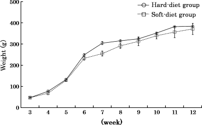
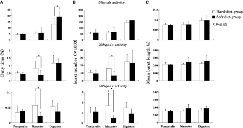
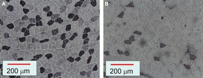
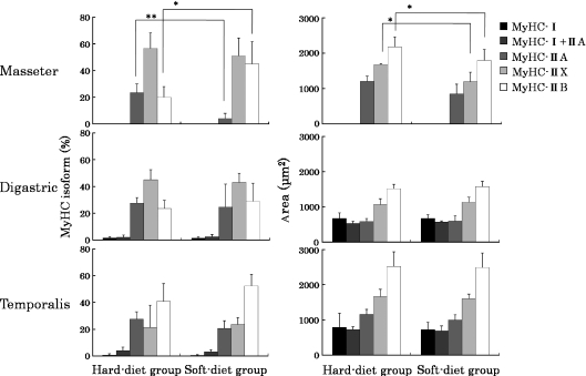
The development of the craniofacial system occurs, among other reasons, as a response to functional needs. In particular, the deficiency of the proper masticatory stimulus affects the growth. The purpose of this study was to relate alterations of muscle activity during postnatal development to adaptational changes in the muscle fibers. Fourteen 21-day-old Wistar strain male rats were randomly divided into two groups and fed on either a solid (hard-diet group) or a powder (soft-diet group) diet for 63 days. A radio-telemetric device was implanted to record muscle activity continuously from the superficial masseter, anterior belly of digastric and anterior temporalis muscles. The degree of daily muscle use was quantified by the total duration of muscle activity per day (duty time), the total burst number and their average length exceeding specified levels of the peak activity (5, 20 and 50%). The fiber type composition of the muscles was examined by the myosin heavy chain content of fibers by means of immunohistochemical staining and their cross-sectional area was measured. All muscle fibers were identified as slow type I and fast type IIA, IIX or IIB (respectively, with increasing twitch contraction speed and fatigability). At lower activity levels (exceeding 5% of the peak activity), the duty time of the anterior belly of the digastric muscle was significantly higher in the soft-diet group than in the hard-diet group (P < 0.05). At higher activity levels (exceeding 20 and 50% of the peak activity), the duty time of the superficial masseter muscle in the soft-diet group was significantly lower than that in the hard-diet group (P < 0.05). There was no difference in the duty time of the anterior temporalis muscle at any muscle activity level. The percentage of type IIA fibers of the superficial masseter muscle in the soft-diet group was significantly lower than that in the hard-diet group (P < 0.01) and the opposite was true with regard to type IIB fibers (P < 0.05). The cross-sectional area of type IIX and type IIB fibers of the superficial masseter muscle was significantly smaller in the soft-diet group than in the hard-diet group (P < 0.05). There was no difference in the muscle fiber composition and the cross-sectional area of the anterior belly of the digastric and anterior temporalis muscles. In conclusion, for the jaw muscles of male rats reared on a soft diet, the slow-to-fast transition of muscle fiber was shown in only the superficial masseter muscle. Therefore, the reduction in the amount of powerful muscle contractions could be important for the slow-to-fast transition of the myosin heavy chain isoform in muscle fibers.
Figures
Similar articles
-
Functional characteristics of the rat jaw muscles: daily muscle activity and fiber type composition.J Anat. 2009 Dec;215(6):656-62. doi: 10.1111/j.1469-7580.2009.01152.x. Epub 2009 Oct 6. J Anat. 2009. PMID: 19811563 Free PMC article.
-
Daily activity of the rabbit jaw muscles during early postnatal development.Neuroscience. 2006 Jun 19;140(1):137-46. doi: 10.1016/j.neuroscience.2006.01.037. Epub 2006 Mar 9. Neuroscience. 2006. PMID: 16529874
-
The masticatory system under varying functional load. Part 1: Structural adaptation of rabbit jaw muscles to reduced masticatory load.Eur J Orthod. 2011 Aug;33(4):359-64. doi: 10.1093/ejo/cjq083. Epub 2010 Oct 5. Eur J Orthod. 2011. PMID: 20923937
-
Muscle mechanics: adaptations with exercise-training.Exerc Sport Sci Rev. 1996;24:427-73. Exerc Sport Sci Rev. 1996. PMID: 8744258 Review.
-
Fiber-type composition of the human jaw muscles--(part 2) role of hybrid fibers and factors responsible for inter-individual variation.J Dent Res. 2005 Sep;84(9):784-93. doi: 10.1177/154405910508400902. J Dent Res. 2005. PMID: 16109985 Review.
Cited by
-
Effects of low occlusal loading on the neuromuscular behavioral development of cortically-elicited jaw movements in growing rats.Sci Rep. 2021 Mar 30;11(1):7175. doi: 10.1038/s41598-021-86581-9. Sci Rep. 2021. PMID: 33785823 Free PMC article.
-
Muscle-Bone Crosstalk in the Masticatory System: From Biomechanical to Molecular Interactions.Front Endocrinol (Lausanne). 2021 Mar 1;11:606947. doi: 10.3389/fendo.2020.606947. eCollection 2020. Front Endocrinol (Lausanne). 2021. PMID: 33732211 Free PMC article. Review.
-
Effects of gum chewing exercise on maximum bite force according to facial morphology.Clin Exp Dent Res. 2018 Feb 22;4(2):48-51. doi: 10.1002/cre2.102. eCollection 2018 Apr. Clin Exp Dent Res. 2018. PMID: 29744215 Free PMC article.
-
Effect of β-hydroxy-β-methylbutyrate in masticatory muscles of rats.J Anat. 2015 Jan;226(1):40-6. doi: 10.1111/joa.12256. Epub 2014 Nov 14. J Anat. 2015. PMID: 25400135 Free PMC article.
-
Intermittent hypoxia inhibits mandibular cartilage growth with reduced TGF-β and SOX9 expressions in neonatal rats.Sci Rep. 2021 Jan 13;11(1):1140. doi: 10.1038/s41598-020-80303-3. Sci Rep. 2021. PMID: 33441835 Free PMC article.
References
-
- Burr DB. Muscle strength, bone mass, and age-related bone loss. J Bone Miner Res. 1997;12:1547–1551. - PubMed
-
- Caiozzo VJ. Plasticity of skeletal muscle phenotype: mechanical consequences. Muscle Nerve. 2002;26:740–768. - PubMed
-
- Delp MD, Pette D. Morphological changes during fiber type transitions in low-frequency-stimulated rat fast-twitch muscle. Cell Tissue Res. 1994;277:363–371. - PubMed
-
- Grossman EJ, Roy RR, Talmadge RJ, et al. Effects of inactivity on myosin heavy chain composition and size of rat soleus fibers. Muscle Nerve. 1998;21:375–389. - PubMed
Publication types
MeSH terms
LinkOut - more resources
Full Text Sources
