. 2010 Jun;60(3):205-17.

Cellular compensatory mechanisms in the CNS of dysmyelinated rats

Affiliations

Cellular compensatory mechanisms in the CNS of dysmyelinated rats

Jacek M Kwiecien. Comp Med. 2010 Jun.

Abstract

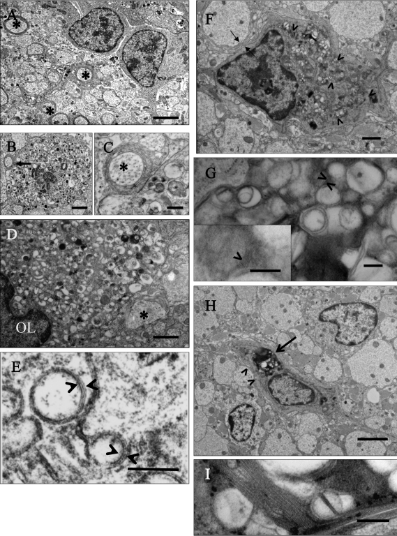
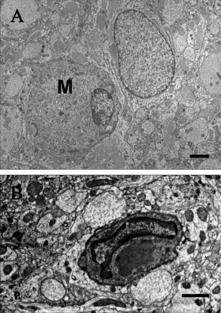
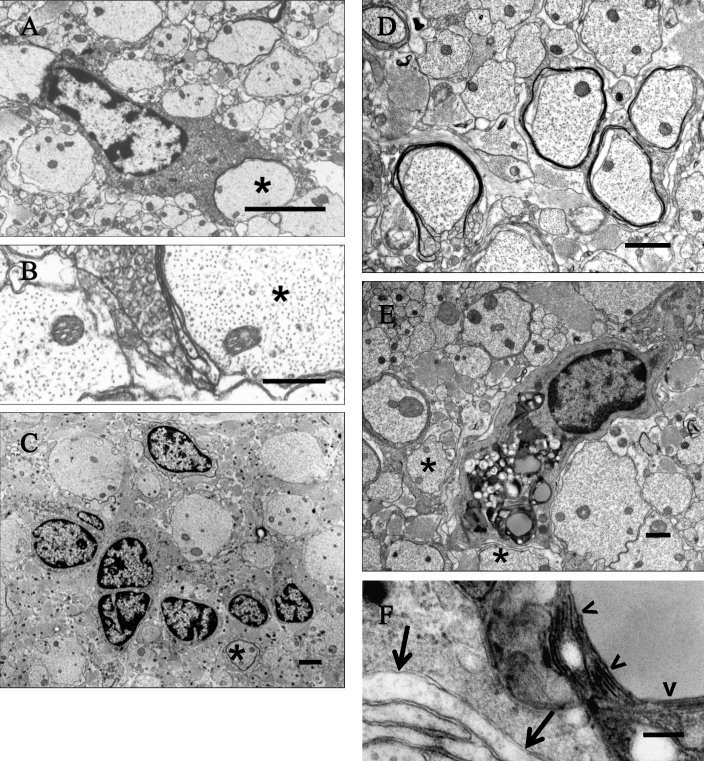
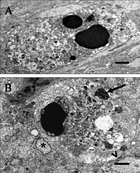
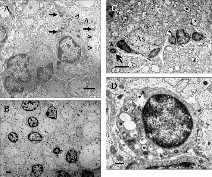
Loss or absolute lack of myelin in the CNS results in remarkable compensation at the cellular level. In this study on the natural progression of neuropathology in the CNS in 2 related but distinct long-lived dysmyelinated rats, total lack of myelin was associated with remarkable glial cell proliferation and ineffective myelinating activity throughout life in Long Evans Bouncer (LE-bo) rats; conversely, in Long Evans Shaker (LES) rats, futile myelinating activity ceased when rats were advanced in age. Progressively severe astrogliosis separates individual axons from each other and coincides with widespread, abundant axonal sprouting throughout the life in both rat strains. Severely dysmyelinated Long Evans rats can serve as excellent models to elucidate the cellular and molecular mechanisms of neuroglial compensation to lack or loss of myelin in vivo and to study axonal plasticity in the adult demyelinated CNS.


Figures

Figure 1.
Figure 1.
Electron micrographs of progression of degeneration of oligodendrocytes in the spinal cord and optic nerve of LES rats 1 to 16 wk old. (A) Spinal cord of a 1-wk-old LES rat, in which 2 glial cells interpreted as immature oligodendrocytes are surrounded by dysmyelinated neuropil with few axons surrounded by thin sheaths (asterisks). Bar, 2 μm. (B) Spinal cord of 1-wk-old LES rat. A degenerating cell is closely associated with a large adjacent axon (arrow) with a few uncompacted lamellae and numerous abnormal vesicular structures in the perikaryon. Bar, 2 μm. (C) Detail of the thinly myelinated axon (asterisk) and degenerating changes in the perikaryon of panel B. Bar, 500 nm. (D) Optic nerve of 4-wk-old LES rat. A degenerating oligodendrocyte is closely adjacent to a large adjacent axon (asterisk). Its perikaryon is enlarged and contains small vesicular structures. Bar, 1 μm. (E) Higher magnification of vesicular structures of panel D, which appear to be limited by a pentalamellar membrane (arrowheads) comprising 3 electrodense lines separated by 2 electrolucent lines with a regular periodicity. Bar, 100 nm. (F) Spinal cord of 8-wk-old LES rat. A degenerating cell forms uncompacted myelin membranes against a large adjacent axon (arrows), and its perikaryon is distended with multiple large aggregations of stacks of membranes or honeycombs (arrowheads). Bar, 1 μm. (G) Higher magnification of panel F showing a fragment of a honeycomb with aggregated vesicular structures. In some areas there are stacks of membranes (arrowheads) that have a regular periodicity of approximately 5 nm between electrodense lines (arrowhead, inset). Bar, 200 nm (inset, 100 nm). (H) Spinal cord of 16-wk-old LES rat. An oligodendrocyte in the center of the image has numerous honeycombs in its perikaryon (arrow) with a few uncompacted myelin lamellae against large adjacent axons (arrowheads). Two adjacent cells appear to be immature oligodendrocytes. Wide astroglial processes separate individual axons from each other. Bar, 2 μm. (I) Detail of a honeycomb with a stack of multiple membranes. Bar, 200 nm.
Figure 2.
Figure 2.
Microglial cells. (A) In this 24-wk-old LES rat, the round cell with a marginalized, elongated nucleus does not form myelin against adjacent axons and lacks vesicular structures and honeycombs in the perikaryon. This cell is considered to be a microglial cell (M) and is adjacent to an astrocyte. Bar, 4 μm. (B) This microglial cell in a 12-wk-old LE-bo rat has a banana-shaped nucleus and a large, amorphous, moderately electrodense inclusion in its cytoplasm. This cell does not form myelin against adjacent axons and lacks vesicular structures or honeycombs in the perikaryon. Bar, 2 μm.
Figure 3.
Figure 3.
Ineffective myelinating activity by oligodendrocytes of the spinal cord and optic nerve of (A through C) adult LES and (D through F) LE-bo rats. (A) LES rat, 16 wk of age. An oligodendrocyte extends its perikaryon in attempt to myelinate a large adjacent axon (asterisk) with an incomplete myelin sheath. Bar, 2 μm. (B) Detail of the incomplete myelin sheath in panel A. Bar, 500 nm. (C) LES rat, 69 wk. A cluster of glial cells interpreted as immature oligodendrocytes is surrounded by naked axons separated from each other by severe astrogliosis, with the exception of one axon that is surrounded by a single lamella (asterisk). Bar, 2 μm. (D) LE-bo rat, 45 wk. Several axons have thin uncompacted and sometimes incomplete sheaths. Bar, 1 μm. (E) LE-bo rat, 45 wk. An oligodendrocyte forms a few incompacted myelin lamellae against adjacent axons (asterisks), and its perikaryon is distended by honeycombs. Bar, 1 μm. (F) Detail of the wall of a honeycomb encompassing stacks of multiple membranes (arrowheads) and of incompacted myelin adjacent to the oligodendrocyte (arrows). Bar, 200 nm.
Figure 4.
Figure 4.
Glial cell death. (A) LES rat, 2 wk of age. An oligodendrocyte with a dense karyorrhectic nucleus and perikaryon distended with vesicular structures. Bar, 2 μm. (B) LE-bo rat, 12 wk. A large oligodendrocyte with a pyknotic nucleus and parikaryon containing scattered vesicles and a large, dense, honeycomb structure. A large axon is separated from the cell membrane by few uncompacted lamellae (open arrowhead); another adjacent axon has a thin uncompacted sheath (asterisk). Bar, 1 μm.
Figure 5.
Figure 5.
Glial cell plasticity in old LES and LE-bo rats. (A) LES rat, 24 wk of age. A mature oligodendrocyte forms incompacted myelin lamellae against its membrane (open arrowheads) and contains numerous honeycombs scattered in the perikaryon (short arrows). Adjacent is a cluster of 4 immature glial cells. Bar, 2 μm. (B) LES rat, 69 wk. Several immature glial cells are scattered among naked axons, which are separated from each other by astrogliosis. Bar, 4 μm. (C) LE-bo rat, 45 wk. A cluster of immature glial cells of oligodendrocyte-type are adjacent to an astrocyte (As). In the surrounding area are scattered axons with thin myelin sheaths (asterisks). The arrow indicates an oligodendrocyte-type cell with multiple vesicles in the perikaryon, which are considered to be degenerative changes. Bar, 4 μm. (D) Area indicated by the arrow in panel C. An oligodendrocyte-type cell with formation of vesicles in the perikaryon, which are considered to be degenerative changes. Bar, 500 nm.
Figure 6.
Figure 6.
Glial cell proliferation in LES and LE-bo rats. (A) Glial cell proliferation in the spinal cord of LES, LE-bo, and LE-control rats (1 through 40 wk of age; n = 4 per group) and single LES (69 wk) and LE-bo (45 wk) rats. For each rat, the numbers of [3H]-thymidine-positive glial cells were counted from 3 consecutive 1-mm-thick sections from the midthoracic spinal cord. Each column represents the mean ± 1 SD of the summaries of counts. The maximum inhibition of glial cell proliferation is at 8 wk for LES rats and at 16 wk for LE-bo rats. (B) Glial cells in a single section of the intracranial optic nerve collected from rats described in panel A. Each column represents the mean ± 1 SD of counts from each rat at the target age.
Figure 7.
Figure 7.
Astrogliosis and axonal plasticity in the dysmyelinated rat CNS. (A) Spinal cord, LES rat, 1 wk of age. An area between 2 oligodendrocytes, with unmyelinated axons closely adjacent to each other except for 2 axons with thin, uncompacted sheaths; one of these axons is adjacent to an oligodendrocyte. Bar 2, μm. (B) Spinal cord, LES rat, 24 wk. Almost all axons in the field adjacent to a hypertrophied astrocyte (As) are separated from each other by astrocytic processes. Bar, 2 μm. (C) Spinal cord, LES rat, 69 wk. Large and medium-sized naked axons (asterisks) are widely separated from each other by astrocytic processes, 1 hypertrophied astrocyte, and 2 immature glial cells, presumably of oligodendroglial lineage. Bar, 4 μm. (D) Optic nerve, LES rat, 16 wk. The cytoplasm of s hypertrophied astrocyte (As) is rich in intermediate filaments (if) and is surrounded by axons, 2 of which have thin incompacted sheaths, but others are naked and sometimes with microtubules-containing sprouts against the astroglial cell membrane. Bar, 1 μm (inset, 200 nm). (E) Optic nerve, LES rat, 16 wk. One large axon (asterisk) adjacent to an oligodendrocyte (OL) has a thin and incomplete sheath; other axons are unmyelinated, are markedly although not completely separated from each other by astrocytic processes, and often have adjacent clusters of small processes with microtubules (sprouts, circled), sometimes adjacent to the cell membrane of an oligodendrocyte. Bar, 1 μm.

Similar articles

Cited by

References

    1. Allen IV, Kirk J. 1992. Demyelinating diseases, p 447–520 In: Adams JH, Duchen LW, Greenfield's neuropathology. New York (NY): Oxford University Press
    1. Baracskay KL, Duchala CS, Miller RH, Macklin WB, Trapp BD. 2002. Oligodedrogenesis is defferentially regulated in gray and white matter of jimpy mice. J Neurosci Res 70:645–654 - PubMed
    1. Barres BA, Raff MC. 1999. Axonal control of oligodendrocyte development. J Cell Biol 147:1123–1128 - PMC - PubMed
    1. Baumann N, Pham-Dinh D. 2001. Biology of oligodendrocyte and myelin in the mammalian central nervous system. Physiol Rev 81:871–927 - PubMed
    1. Bradl M, Linington C. 1996. Animal models of demyelination. Brain Pathol 6:303–311 - PMC - PubMed

Publication types

MeSH terms

LinkOut - more resources