. 2010 Jul 9;87(1):26-39.
doi: 10.1016/j.ajhg.2010.05.019. Epub 2010 Jun 24.

X-linked cone dystrophy caused by mutation of the red and green cone opsins

Affiliations

X-linked cone dystrophy caused by mutation of the red and green cone opsins

Jessica C Gardner et al. Am J Hum Genet. .

Abstract

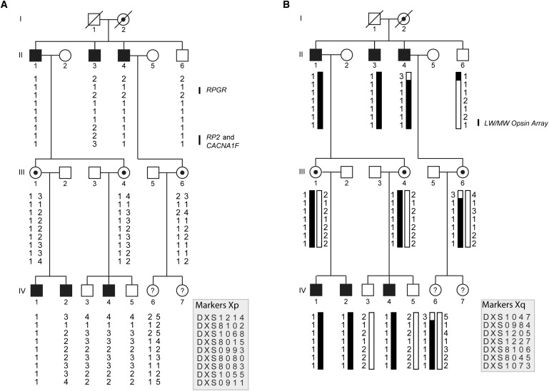
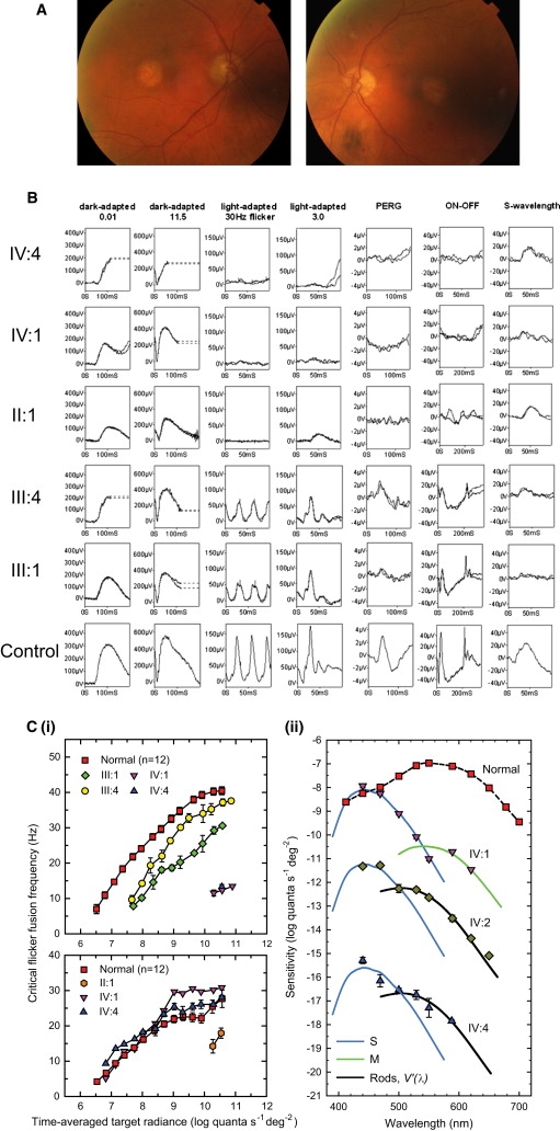
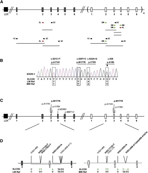
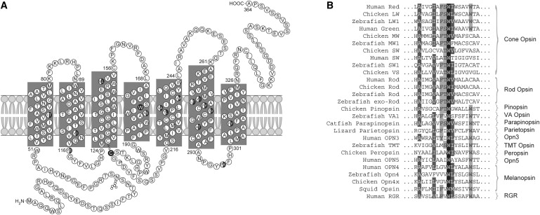
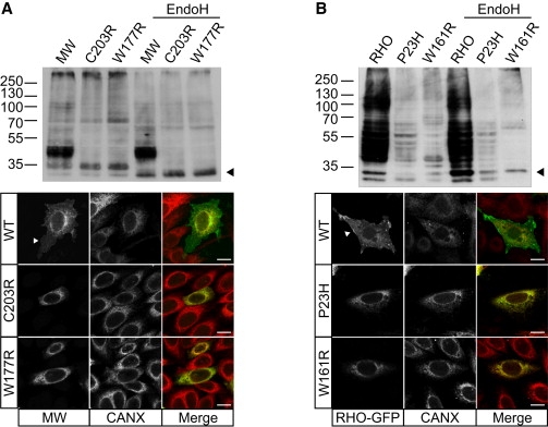
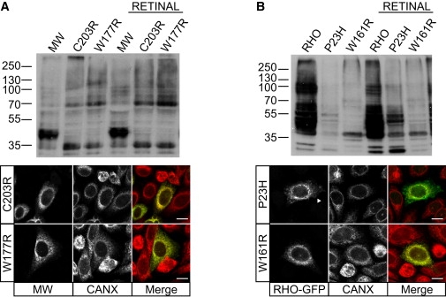
X-linked cone and cone-rod dystrophies (XLCOD and XLCORD) are a heterogeneous group of progressive disorders that solely or primarily affect cone photoreceptors. Mutations in exon ORF15 of the RPGR gene are the most common underlying cause. In a previous study, we excluded RPGR exon ORF15 in some families with XLCOD. Here, we report genetic mapping of XLCOD to Xq26.1-qter. A significant LOD score was detected with marker DXS8045 (Z(max) = 2.41 [theta = 0.0]). The disease locus encompasses the cone opsin gene array on Xq28. Analysis of the array revealed a missense mutation (c. 529T>C [p. W177R]) in exon 3 of both the long-wavelength-sensitive (LW, red) and medium-wavelength-sensitive (MW, green) cone opsin genes that segregated with disease. Both exon 3 sequences were identical and were derived from the MW gene as a result of gene conversion. The amino acid W177 is highly conserved in visual and nonvisual opsins across species. We show that W177R in MW opsin and the equivalent W161R mutation in rod opsin result in protein misfolding and retention in the endoplasmic reticulum. We also demonstrate that W177R misfolding, unlike the P23H mutation in rod opsin that causes retinitis pigmentosa, is not rescued by treatment with the pharmacological chaperone 9-cis-retinal. Mutations in the LW/MW cone opsin gene array can, therefore, lead to a spectrum of disease, ranging from color blindness to progressive cone dystrophy (XLCOD5).


Figures

Figure 1
Figure 1
Pedigree and Haplotype Analysis of the XLCOD Family, Defining the Critical Interval as Xq26.1-qter (A) Haplotypes for markers on Xp show that the disease does not segregate with known disease genes (RPGR, RP2, and CACNA1F). (B) XLCOD segregates with markers on Xq26.1-qter. The affected haplotype is shaded black. Individuals II:4, II:6, and III:6 have a recombination between markers DXS1047 and DXS984, defining the proximal boundary of disease. No distal recombinations were identified; thus, the critical interval is defined as DXS1047-Xqter. The location of the LW/MW opsin array is indicated. Haplotypes are shown according to marker order on the X chromosome, as indicated in the key. Filled symbol, affected; empty symbol, unaffected; circles with dots, obligate carrier females; ?, affectation status unknown.
Figure 2
Figure 2
Phenotype of the XLCOD5 Family (A) Color fundus photographs of affected male patient II:3 (80 yrs old), showing symmetrical bilateral macular atrophy. (B) PERG and full-field ERGs for affected males IV:4, IV:1, and II:1 and obligate carrier females III:1 and III:4, as indicated, compared with those from a representative normal subject (bottom row). Recordings were performed with gold-foil corneal electrodes. Broken lines replace eye-movement artifacts. (C) L-cone cff measurements (i, upper panel) for two affected males (IV:1 and IV:4) and two obligate carriers (III:1 and III:4), showing significant loss of L- and M-cone sensitivity in affected males and reduced L-cone sensitivity in both carriers as compared to controls. S-cone critical flicker fusion measurements (i, lower panel) for three affected males (IV:1, IV:4, and II:1), showing significant S-cone sensitivity loss in the eldest affected male (II:1) as compared to controls. Spectral-sensitivity measurements (ii) for the three youngest affected males (IV:1, IV:2, and IV:4), showing substantial losses of flicker sensitivity at middle and long wavelengths, but sensitivities at short wavelengths that are nearly normal. Error bars represent ±1 SEM.
Figure 3
Figure 3
Molecular Analysis of the Cone Opsin Array and Identification of a c.529T>C Mutation in Exon 3 of the LW and MW Cone Opsin Genes (A) The cone opsin array comprises an upstream LCR, an LW opsin gene, and one or more MW opsin genes lying in a 5′ to 3′ position in tandem on Xq28. Grey boxes represent LW opsin exons, and white boxes represent MW opsin exons. Exons are numbered below the figure. A subscript “n” represents one or more MW opsin genes. The black box represents the upstream LCR. After an initial screen of the LCR and coamplification of exons of the LW/MW genes, a strategy was developed to determine the precise opsin array structure in the XLCOD5 family. A combination of gene-specific primers (labeled “L” or “M”) and coamplification primers (labeled “C”) was used, depicted beneath the diagrammatical representation of the opsin array. (B) Electropherogram of exon 3 in an affected male (IV:2). Reference sequences of both LW and MW genes are shown beneath the patient electropherogram (XLCOD). The dotted boxes represent known polymorphic nucleotide variants, with the resulting amino acid variant shown above. The lined box highlights the location of a c.529T>C missense mutation in exon 3 resulting in p.W177R. The arrow over a dotted box highlights a single G peak representing sequence derived from exon 3 of an MW gene only (p. A180). This indicates that the sequence changes shown are derived from an MW exon 3 only. Electropherograms for other exons (1, 2, 4, 5, and 6) in the affected male (IV:2) are shown in Figure S1. (C) Diagrammatic representation of the cone opsin array in the XLCOD family. Both LW and MW opsin genes contain identical copies of mutant MW exon 3 with the c.529T>C missense mutation (p.W177R). Polymorphic and nonsynonymous substitutions are also indicated (italic). (D) Detailed expansion of sequence between exons 2 and 4 of the LW and MW opsin genes in an affected male (XLCOD) compared to reference sequences. Identified nucleotide substitutions facilitated further characterization of the gene-conversion event leading to a mutated MW exon 3 in both the LW and MW genes in the opsin array. Dotted line indicates MW-gene-derived sequence, solid line indicates LW-gene-derived sequence. The upper and lower dotted lines in the LW opsin gene represent the minimal and maximal gene-conversion tracts, respectively. IVS2-171G>T and IVS3+25G>A present in the LW gene are derived from the MW gene, and they define the minimal gene-conversion interval surrounding the mutated MW exon 3.
Figure 4
Figure 4
Secondary Structure of the Cone Opsins and Conservation of W177 (A) The secondary structure of the red (LW) and green (MW) cone opsins. Amino acid differences between the LW and MW opsins are shown as half-closed circles. W177 in transmembrane domain 4 and C203 are shown as a closed circle. (B) Sequence alignment encompassing W177 of LW and MW opsins with known visual and nonvisual opsins in a variety of species, as named on the left. This tryptophan is 100% conserved in all opsins.
Figure 5
Figure 5
The W177R MW Mutation Causes a Cone Opsin Misfolding Defect Resulting in ER Retention (A) Immunoblot analysis of SK-N-SH cell lysates (10 ug DM-soluble lysate) transfected with either WT MW opsin (MW), C203R MW mutant or W177R MW mutant, with Endo H treatment as indicated, probed with 1D4 opsin antibody. Both W177R and C203R show fewer glycosylated species as compared to WT and a shift in glycosylated species following Endo H treatment (arrowhead). Expression of WT MW opsin (top), C203R (middle), or W177R (bottom) in SK-N-SH cells detected with 1D4 (green in merged panel) and counterstained for the ER marker calnexin (Cnx, red in merged panel). WT cone opsin (MW) is processed in the ER and targeted to the plasma membrane (arrowhead), reflecting normal biogenesis and traffic of MW opsin, but both mutants were retained in the ER and colocalized with calnexin. (B) Immunoblot analysis of SK-N-SH cell lysates transfected with WT rod opsin (RHO) and the P23H and W161R RHO mutants, with Endo H treatment as indicated, probed with 1D4 antibody. Endo H treatment results in a shift in glycosylated species of the W161R mutant (arrowhead). Expression of GFP-tagged RHO (top), P23H (middle), and W161R (bottom) (green) counterstained for calnexin (red). RHO-GFP is detected at the plasma membrane, but both mutant proteins are retained in the ER.
Figure 6
Figure 6
The W177R MW Mutation Is Not Rescued with 9-cis-Retinal (A) Immunoblot analysis of SK-N-SH cell lysates transfected with WT MW opsin, W177R MW mutant and C203R MW mutant opsins, with and without 9-cis-retinal treatment, as indicated. 9-cis-retinal treatment had no effect on the level or SDS-PAGE mobility of either of the MW mutants. Immunofluorescence confirmed no retinoid-associated changes in the intracellular traffic of the mutants. (B) Immunoblot analysis of SK-N-SH cell lysates transfected with RHO and the RHO mutants P23H and W161R, with and without 9-cis-retinal treatment resulted in increased expression of P23H, but not W161R. 9-cis-retinal treatment led to an increase in P23H traffic to the plasma membrane (arrowhead), whereas W161R was retained in the ER, where it colocalized with calnexin.

References

    1. Michaelides M., Hardcastle A.J., Hunt D.M., Moore A.T. Progressive cone and cone-rod dystrophies: phenotypes and underlying molecular genetic basis. Surv. Ophthalmol. 2006;51:232–258. - PubMed
    1. Demirci F.Y., Rigatti B.W., Wen G., Radak A.L., Mah T.S., Baic C.L., Traboulsi E.I., Alitalo T., Ramser J., Gorin M.B. X-linked cone-rod dystrophy (locus COD1): identification of mutations in RPGR exon ORF15. Am. J. Hum. Genet. 2002;70:1049–1053. - PMC - PubMed
    1. Yang Z., Peachey N.S., Moshfeghi D.M., Thirumalaichary S., Chorich L., Shugart Y.Y., Fan K., Zhang K. Mutations in the RPGR gene cause X-linked cone dystrophy. Hum. Mol. Genet. 2002;11:605–611. - PubMed
    1. Ebenezer N.D., Michaelides M., Jenkins S.A., Audo I., Webster A.R., Cheetham M.E., Stockman A., Maher E.R., Ainsworth J.R., Yates J.R. Identification of novel RPGR ORF15 mutations in X-linked progressive cone-rod dystrophy (XLCORD) families. Invest. Ophthalmol. Vis. Sci. 2005;46:1891–1898. - PubMed
    1. Ruddle J.B., Ebenezer N.D., Kearns L.S., Mulhall L.E., Mackey D.A., Hardcastle A.J. RPGR ORF15 genotype and clinical variability of retinal degeneration in an Australian population. Br. J. Ophthalmol. 2009;93:1151–1154. - PubMed

Publication types

MeSH terms