

Anatomic-manometric correlation of the upper esophageal sphincter: a concurrent US and manometry study
- PMID: 20579650
- PMCID: PMC4142490
- DOI: 10.1016/j.gie.2010.04.029
Anatomic-manometric correlation of the upper esophageal sphincter: a concurrent US and manometry study
Abstract
Background: The pharyngoesophageal segment commonly referred to as the upper esophageal sphincter (UES) generates a high-pressure zone (HPZ) between the pharynx and the esophagus. However, the exact anatomical components of the UES-HPZ remain incompletely determined.
Objective: To systematically define the US signature of various components of the pharyngoesophageal junction and to determine how these structures contribute to the development of the UES-HPZ.
Design: Prospective, experimental study.
Setting: Tertiary Academic Medical Center.
Patients: This study involved 18 healthy volunteers.
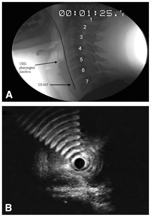
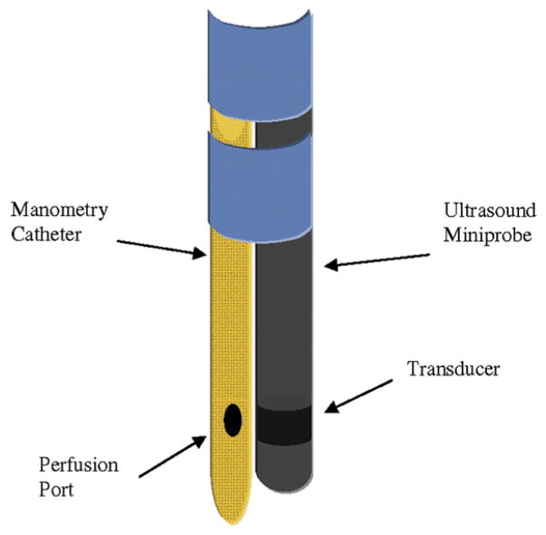
Intervention: We studied 5 participants by using a high-frequency US miniprobe (US-MP) and concurrent fluoroscopy and another 13 participants by using the US-MP and concurrent manometry.
Main outcome measurements: Relative contribution of various muscles in the UES-HPZ.
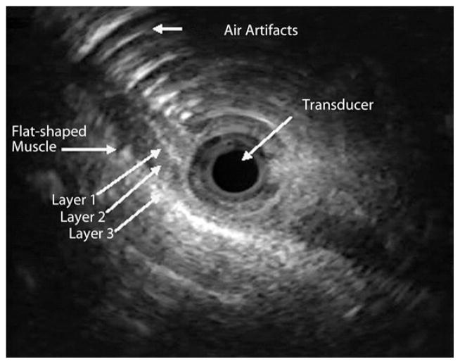
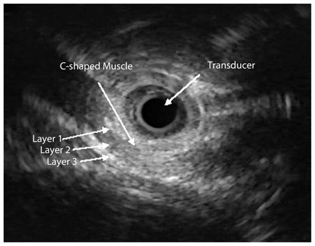
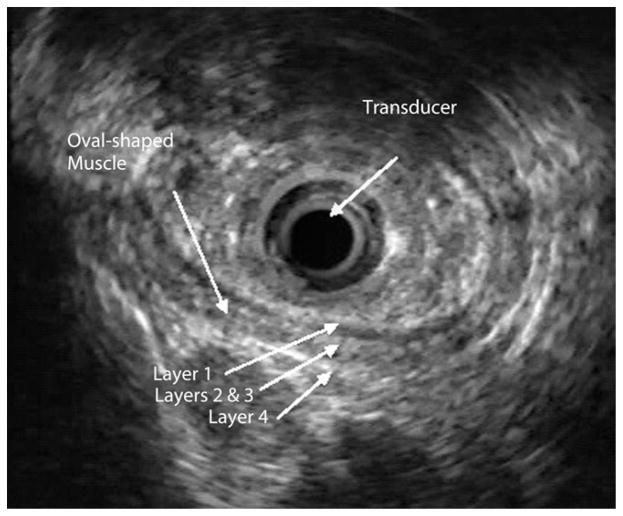
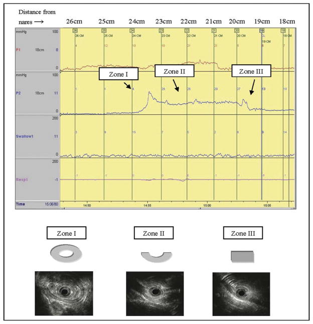
Results: Manometrically, the UES-HPZ had a median length of 4.0 cm (range 3.0-4.5 cm). A C-shaped muscle, believed to represent the cricopharyngeus muscle, was observed for a median length of 3.5 cm (range 2.0-4.0 cm). The oval configuration representing the esophageal contribution to the UES was seen in 10 of 13 participants (77%) at the distal HPZ (esophagus to UES transition zone). The flat configuration of the inferior constrictor muscle was noted in 7 of 13 participants (54%) at the proximal HPZ (UES to pharynx transition zone). There were 4 to 5 wall layers versus 3 layers in the distal and proximal HPZ, respectively. The mean (+/- SD) muscle thickness was relatively constant along the length of the UES-HPZ.
Limitations: Air artifacts in the UES-HPZ.
Conclusion: The configuration and layers of the UES-HPZ vary along its length. The upper esophagus is a significant contributor to the distal UES-HPZ.
Copyright 2010 American Society for Gastrointestinal Endoscopy. Published by Mosby, Inc. All rights reserved.
Figures
References
-
- Liu JB, Miller LS, Goldberg BB, et al. Transnasal US of the esophagus: preliminary morphologic and function studies. Radiology. 1992;184:721–7. - PubMed
-
- Shaker R, Ren J, Xie P, et al. Characterization of the pharyngo-UES contractile reflex in humans. Am J Physiol. 1997;273:G854–8. - PubMed
-
- Mittal RK, Liu J, Puckett JL, et al. Sensory and motor function of the esophagus: lessons from ultrasound imaging. Gastroenterology. 2005;128:487–97. - PubMed
-
- Chak A, Soweid A, Hoffman B, et al. Clinical implications of catheter probe-assisted endoluminal ultrasonography. Endoscopy. 1998;30:A169–72. - PubMed
-
- Menzel J, Domschke W. Gastrointestinal miniprobe sonography: the current status. Am J Gastroenterol. 2000;95:605–16. - PubMed
Publication types
MeSH terms
Grants and funding
LinkOut - more resources
Full Text Sources
Other Literature Sources
