. 2010 Sep 15;405(1):139-48.
doi: 10.1016/j.virol.2010.05.031. Epub 2010 Jul 1.

Palmitoylation of SARS-CoV S protein is necessary for partitioning into detergent-resistant membranes and cell-cell fusion but not interaction with M protein

Affiliations

Palmitoylation of SARS-CoV S protein is necessary for partitioning into detergent-resistant membranes and cell-cell fusion but not interaction with M protein

Corrin E McBride et al. Virology. .

Abstract

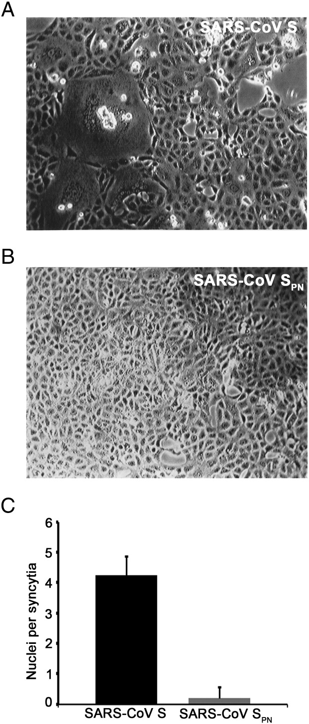
Coronaviruses are enveloped RNA viruses that generally cause mild disease in humans. However, the recently emerged coronavirus that caused severe acute respiratory syndrome (SARS-CoV) is the most pathogenic human coronavirus discovered to date. The SARS-CoV spike (S) protein mediates virus entry by binding cellular receptors and inducing fusion between the viral envelope and the host cell membrane. Coronavirus S proteins are palmitoylated, which may affect function. Here, we created a non-palmitoylated SARS-CoV S protein by mutating all nine cytoplasmic cysteine residues. Palmitoylation of SARS-CoV S was required for partitioning into detergent-resistant membranes and for cell-cell fusion. Surprisingly, however, palmitoylation of S was not required for interaction with SARS-CoV M protein. This contrasts with the requirement for palmitoylation of mouse hepatitis virus S protein for interaction with M protein and may point to important differences in assembly and infectivity of these two coronaviruses.


Figures

Fig. 1
Fig. 1
SARS-CoV S palmitoylation can occur in a pre-medial Golgi compartment and is a stable modification. (A) At 24 h post-transfection, HEK293T cells expressing SARS-CoV S were labeled with 35S-methionine/cysteine or 3H-palmitic acid for 30 min. After lysis, S protein was immunoprecipitated, denatured, digested with endo H, separated by SDS–PAGE and imaged by fluorography. (B) At 24 h post-transfection, HEK293T cells expressing SARS-CoV S were labeled with 35S-methionine/cysteine or 3H-palmitic acid for 30 min and chased for 0, 40, 80 or 120 min. After lysis, S protein was immunoprecipitated, separated by SDS–PAGE and analyzed by fluorography for 24 h (35S) or for 1 wk (3H).
Fig. 2
Fig. 2
SARS-CoV S lacking all cytoplasmic cysteines is not palmitoylated. (A) Cytoplasmic tail sequence of SARS-CoV S and a mutant lacking all 9 cytoplasmic cysteines (SARS-CoV SPN). (B) At 24 h post-transfection, HEK293T cells expressing SARS-CoV S or SPN were labeled with 35S-methionine/cysteine or 3H-palmitic acid for 30 min. After lysis, S protein was immunoprecipitated, separated by SDS–PAGE and analyzed by fluorography. Longer exposure (6 weeks) of the 3H-palmitate labeled samples confirmed the absence of palmitoylated SARS-CoV SPN.
Fig. 3
Fig. 3
SARS-CoV S and SARS-CoV SPN have similar half-lives. At 24 h post-transfection, HEK293T cells expressing SARS-CoV S (solid line) or SPN (dashed line) were labeled with 35S-methionine/cysteine for 20 min and chased for 0, 1, 2 or 3 h. After lysis, S protein was immunoprecipitated, separated by SDS–PAGE and analyzed by autoradiography. Percentage of S or SPN remaining at each time point was calculated using the amount of S or SPN at 0 h chase as 100%. The average of 3 independent experiments ± SEM is shown.
Fig. 4
Fig. 4
SARS-CoV S and SARS-CoV SPN both localize to the cell surface. (A) At 24 h post-transfection, HEK293T expressing SARS-CoV S or SPN were fixed, permeabilized and co-stained with mouse anti-SARS-CoV S and rabbit anti-golgin 160 (a Golgi marker). (B) At 24 h post-transfection, unpermeabilized HEK293T cells were stained with mouse anti-SARS-CoV S at 0 °C for 20 min to label SARS-CoV S or SPN present on the cell surface. Cells were then fixed, permeabilized and co-stained with rabbit anti-SARS-CoV S to label total S protein. (A and B) Secondary antibodies were Alexa 488-conjugated donkey anti-mouse IgG and Texas Red-conjugated donkey anti-rabbit IgG. The same field is shown in each set of images. (C) At 24 h post-transfection, SARS-CoV S or SPN present at the plasma membrane was biotinylated with a membrane-impermeable biotinylating agent. After lysis, biotinylated S proteins were recovered with streptavidin agarose and analyzed by SDS–PAGE and Western blotting with rabbit anti-SARS-CoV S. The graph shows quantification from 3 independent experiments ± SEM.
Fig. 5
Fig. 5
SARS-CoV S and SARS-CoV SPN can be retained at the Golgi by SARS-CoV M. (A) At 24 h post-transfection, HEK293T cells expressing SARS-CoV S or SPN and SARS-CoV M were fixed, permeabilized and co-stained with mouse anti-SARS-CoV and rabbit anti-SARS-CoV M. Secondary antibodies were Alexa 488-conjugated donkey anti-mouse IgG and Texas Red-conjugated donkey anti-rabbit IgG. (B) At 24 h post-transfection, HEK293T cells expressing SARS-CoV S or SARS-CoV SPN were labeled with 35S-methionine/cysteine for 20 min and chased for 0, 20 or 40 min. After lysis, S protein was immunoprecipitated, denatured, digested with endo H, separated by SDS–PAGE and analyzed by autoradiography. The average of 3 independent experiments ± SEM is shown.
Fig. 6
Fig. 6
SARS-CoV S palmitoylation is necessary for partitioning into detergent-resistant membranes. At 24 h post-transfection, detergent-resistant membranes (DRMs) were extracted from HEK293T cells expressing SARS-CoV S or SPN using cold Triton. DRMs were isolated using discontinuous density ultracentrifugation, and fractions were collected from the top. S protein was identified by Western blotting (upper and middle panels) and DRMs were identified using HRP-cholera toxin B, which binds ganglioside GM1 (bottom panel). A representative image of 3 independent experiments is shown.
Fig. 7
Fig. 7
SARS-CoV S palmitoylation is necessary for cell–cell fusion. At 48 h post-transfection, Vero cells expressing SARS-CoV S (A) or SARS-CoV SPN (B) were trypsinized then re-plated. At 24 h post-trypsinization, the number of nuclei per syncytia was counted; the average of 3 independent experiments ± SEM is shown (C).

References

    1. Baker T.L., Zheng H., Walker J., Coloff J.L., Buss J.E. Distinct rates of palmitate turnover on membrane-bound cellular and oncogenic H-ras. J. Biol. Chem. 2003;278(21):19292–19300. - PubMed
    1. Bisht H., Roberts A., Vogel L., Bukreyev A., Collins P.L., Murphy B.R., Subbarao K., Moss B. Severe acute respiratory syndrome coronavirus spike protein expressed by attenuated vaccinia virus protectively immunizes mice. Proc. Natl. Acad. Sci. U. S. A. 2004;101(17):6641–6646. - PMC - PubMed
    1. Bos E.C., Heijnen L., Luytjes W., Spaan W.J. Mutational analysis of the murine coronavirus spike protein: effect on cell-to-cell fusion. Virology. 1995;214(2):453–463. - PMC - PubMed
    1. Boscarino J.A., Logan H.L., Lacny J.J., Gallagher T.M. Envelope protein palmitoylations are crucial for murine coronavirus assembly. J. Virol. 2008;82(6):2989–2999. - PMC - PubMed
    1. Bosch B.J., de Haan C.A., Smits S.L., Rottier P.J. Spike protein assembly into the coronavirion: exploring the limits of its sequence requirements. Virology. 2005;334(2):306–318. - PMC - PubMed

Publication types

MeSH terms