Comparative Study
. 2011 Dec;32(12):2321.e13-23.
doi: 10.1016/j.neurobiolaging.2010.05.011. Epub 2010 Jun 26.

Age-related neuronal loss in the cochlea is not delayed by synaptic modulation

Affiliations
Comparative Study

Age-related neuronal loss in the cochlea is not delayed by synaptic modulation

David Jin et al. Neurobiol Aging. 2011 Dec.

Abstract

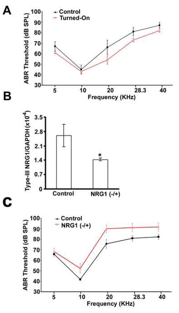
Age-related synaptic change is associated with the functional decline of the nervous system. It is unknown whether this synaptic change is the cause or the consequence of neuronal cell loss. We have addressed this question by examining mice genetically engineered to over- or underexpress neuregulin-1 (NRG1), a direct modulator of synaptic transmission. Transgenic mice overexpressing NRG1 in spiral ganglion neurons (SGNs) showed improvements in hearing thresholds, whereas NRG1 -/+ mice show a complementary worsening of thresholds. However, no significant change in age-related loss of SGNs in either NRG1 -/+ mice or mice overexpressing NRG1 was observed, while a negative association between NRG1 expression level and survival of inner hair cells during aging was observed. Subsequent studies provided evidence that modulating NRG1 levels changes synaptic transmission between SGNs and hair cells. One of the most dramatic examples of this was the reversal of lower hearing thresholds by "turning-off" NRG1 overexpression. These data demonstrate for the first time that synaptic modulation is unable to prevent age-related neuronal loss in the cochlea.


Figures

Fig. 1
Fig. 1
Validation of NRG1 conditionally tissue-specific transgenic mice. (A) Schematic diagram of the conditionally tissue-specific transgenic model, which resulted from a crossing of two transgenic mouse lines. One line had the tTA expression under the CamKIIα promoter, and another line we made contained the biTet-O promoter controlling the expression of both the reporter gene LacZ and the transgene NRG1-EGFP. (B) The LacZ staining showed that SGNs were the only cell type expressing the LacZ. (C) The SGN-specific LacZ expression could be turned-on or off by dox. (D) and (E) Real-time RT-PCR quantifications of the transgene expression (with EGFP primers) or the total expression of NRG1 in the cochlea (n=4, t-test, p < 0.05).
Fig. 2
Fig. 2
No changes in the expression of NRG1 isoforms or their receptors after type-III NRG1 over-expression. (A) RT-PCR analysis of type-I, -II, -III NRG1, and ErbB 2, 3, 4 in cochleae from mice over-expressing type-III NRG1 or their sibling mice without NRG1 transgene at four months of age. No dramatic difference between the control and NRG1 over-expressing mice was observed. (B) Real-time RT-PCR quantification of the same groups of gene expression in the cochlea. No significant difference was found between the control and mice over-expressing NRG1 for NRG1 isoforms and their receptors except type-III NRG1 (n=4, t-test).
Fig. 3
Fig. 3
The NRG1 level modulates ABR thresholds. (A) ABR thresholds (Mean ±S.D) for mice over-expressing NRG1 (red) and their sibling mice without NRG1 transgene (Control; black) at 12 months of age (n=5 for each group, two females and three males; ANOVA, p < 0.05). (B) Real-time RT-PCR quantification of type-III NRG1 in mouse cochleae for the control and NRG1 −/+ mice at four months old (n=4; t-test, p < 0.05). (C) ABR thresholds (Mean ±S.D) for the control (n=8) and NRG1 −/+ mice (n=9) at 12 months old (ANOVA, p < 0.05).
Fig. 4
Fig. 4
Quantitative comparisons of SGNs, OHCs, and IHCs between the control and NRG1 over-expression mice, or the control and NRG1 −/+ Mice. (A). Quantitative comparison of total SGN number among the same four groups of animals tested in Figure 3. No significant difference was detected between the control and transgenic mice at 12 months old mice (t-test). (B) Quantitative comparison of missing OHCs at the 40–70% region from the apex (two-way ANOVA). (C) Quantitative comparison of missing IHCs in the same area (two-way ANOVA).
Fig. 5
Fig. 5
Quantitative comparisons of the expression of two synaptic genes during aging between the control and NRG1 over-expression mice. Age-related up-regulation of PSD-95 (A) and SNAP-25 (B) was found in the cochlea, and this significant up-regulation was diminished for these two genes after NRG1 over-expression (n=4, t-test).
Fig. 6
Fig. 6
Comparisons of ABR hearing thresholds between the control and NRG1 over-expression mice during aging. ABR threshold shifts (Mean ±S.D) for both the control and NRG1 over-expression mice were measured at 5, 10, 20, 28.3, and 40 kHz (n=9 for each group, five females and four males). The dotted line for each group represents the average threshold from these five measurement. The average thresholds increased during for both group, but the differences in average threshold between the two groups remained almost the same across ages, around 5 dB.
Fig. 7
Fig. 7
Comparisons of the input-output curves of Wave I between the control and NRG1 transgenic mice. (A) At two months old, there was no difference between these two groups under C57/6J genetic background in their input-out curves (n=5; two-way ANOVA). (B) At 12 months old, the input-output curve was much higher for NRG1 over-expression group. Please note that the amplitude at the same input intensity was much lower compared the amplitude from two months old mice (n=5; two-way ANOVA, p < 0.001). (C) At 12 months old, the input-out curve was lower at the high intensity range (over 81 dB) for the NRG1 −/+ mice, which expressed a lower amount of NRG1 (n=8; two-way ANOVA, p < 0.001).
Fig. 8
Fig. 8
Reversibility of improved ABR thresholds. ABR thresholds (Mean ±S.D) were measured at four months old for the group with NRG1 over-expression always “turned-off” (n=7; solid black), or the group with NRG1 over-expression “turned-on” at two months old (n-9; solid gray). ABR thresholds (Mean ±S.D) were measured again for the late group after NRG1 over-expression was subsequently “turned-off” for two weeks.

Similar articles

Cited by

References

    1. Adlkofer K, Lai C. Role of neuregulins in glial cell development. Glia. 2000;29:104–111. - PubMed
    1. Bao J. Signal transduction by the cytoplasmic domain of Neuregulin-1 and its roles in neuronal aging. Curr. Signal. Trans. Therapy. 2007;2:240–245.
    1. Bao J, Lei D, Du Y, Ohlemiller KK, Beaudet AL, Role LW. Requirement of nicotinic acetylcholine receptor subunit beta2 in the maintenance of spiral ganglion neurons during aging. J. Neurosci. 2005;25:3041–3045. - PMC - PubMed
    1. Bao J, Lin H, Ouyang Y, Lei D, Osman A, Kim TW, Mei L, Dai P, Ohlemiller KK, Ambron RT. Activity-dependent transcription regulation of PSD-95 by neuregulin-1 and Eos. Nat. Neurosci. 2004;7:1250–1258. - PubMed
    1. Bao J, Wolpowitz D, Role LW, Talmage DA. Back signaling by the Nrg-1 intracellular domain. J. Cell. Biol. 2003;161:1133–1141. - PMC - PubMed

Publication types

MeSH terms