

Complete genome sequence analysis of candidate human rotavirus vaccine strains RV3 and 116E
- PMID: 20580391
- PMCID: PMC2925181
- DOI: 10.1016/j.virol.2010.06.005
Complete genome sequence analysis of candidate human rotavirus vaccine strains RV3 and 116E
Abstract
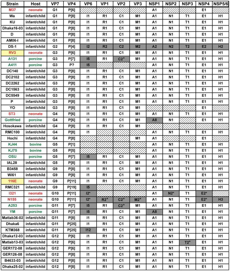
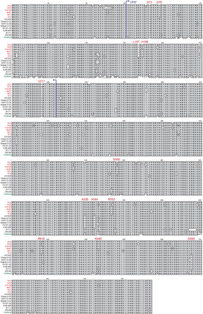
Rotaviruses (RVs) cause severe gastroenteritis in infants and young children; yet, several strains have been isolated from newborns showing no signs of clinical illness. Two of these neonatal strains, RV3 (G3P[6]) and 116E (G9P[11]), are currently being developed as live-attenuated vaccines. In this study, we sequenced the eleven-segmented double-stranded RNA genomes of cell culture-adapted RV3 and 116E and compared their genes and protein products to those of other RVs. Using amino acid alignments and structural predictions, we identified residues of RV3 or 116E that may contribute to attenuation or influence vaccine efficacy. We also discovered residues of the VP4 attachment protein that correlate with the capacity of some P[6] strains, including RV3, to infect newborns versus older infants. The results of this study enhance our understanding of the molecular determinants of RV3 and 116E attenuation and are expected to aid in the ongoing development of these vaccine candidates.
Published by Elsevier Inc.
Figures


Similar articles
-
Molecular characterisation of rotavirus strains detected during a clinical trial of the human neonatal rotavirus vaccine (RV3-BB) in Indonesia.Vaccine. 2018 Sep 18;36(39):5872-5878. doi: 10.1016/j.vaccine.2018.08.027. Epub 2018 Aug 23. Vaccine. 2018. PMID: 30145099 Free PMC article. Clinical Trial.
-
Diversity of rotavirus strains circulating in Northern Brazil after introduction of a rotavirus vaccine: high prevalence of G3P[6] genotype.J Med Virol. 2014 Jun;86(6):1065-72. doi: 10.1002/jmv.23797. Epub 2013 Oct 17. J Med Virol. 2014. PMID: 24136444
-
Reverse Genetics Approach for Developing Rotavirus Vaccine Candidates Carrying VP4 and VP7 Genes Cloned from Clinical Isolates of Human Rotavirus.J Virol. 2020 Dec 22;95(2):e01374-20. doi: 10.1128/JVI.01374-20. Print 2020 Dec 22. J Virol. 2020. PMID: 33087468 Free PMC article.
-
Circulating rotavirus strains in children with acute gastroenteritis in Iran, 1986 to 2023 and their genetic/antigenic divergence compared to approved vaccines strains (Rotarix, RotaTeq, ROTAVAC, ROTASIIL) before mass vaccination: Clues for vaccination policy makers.Virus Res. 2024 Aug;346:199411. doi: 10.1016/j.virusres.2024.199411. Epub 2024 Jun 3. Virus Res. 2024. PMID: 38823689 Free PMC article. Review.
-
Genetic diversity of G1P[8] rotavirus VP7 and VP8* antigens in Finland over a 20-year period: No evidence for selection pressure by universal mass vaccination with RotaTeq® vaccine.Infect Genet Evol. 2013 Oct;19:51-8. doi: 10.1016/j.meegid.2013.06.026. Epub 2013 Jul 4. Infect Genet Evol. 2013. PMID: 23831933 Review.
Cited by
-
Combinatorial tetramer staining and mass cytometry analysis facilitate T-cell epitope mapping and characterization.Nat Biotechnol. 2013 Jul;31(7):623-9. doi: 10.1038/nbt.2593. Epub 2013 Jun 9. Nat Biotechnol. 2013. PMID: 23748502 Free PMC article.
-
Absence of genetic differences among G10P[11] rotaviruses associated with asymptomatic and symptomatic neonatal infections in Vellore, India.J Virol. 2014 Aug;88(16):9060-71. doi: 10.1128/JVI.01417-14. Epub 2014 Jun 4. J Virol. 2014. PMID: 24899175 Free PMC article.
-
Development of improved vaccine cell lines against rotavirus.Sci Data. 2017 Mar 1;4:170021. doi: 10.1038/sdata.2017.21. Sci Data. 2017. PMID: 28248921 Free PMC article.
-
Human Neonatal Rotavirus Vaccine (RV3-BB) to Target Rotavirus from Birth.N Engl J Med. 2018 Feb 22;378(8):719-730. doi: 10.1056/NEJMoa1706804. N Engl J Med. 2018. PMID: 29466164 Free PMC article. Clinical Trial.
-
Isolation and characterization of a new candidate human inactivated rotavirus vaccine strain from hospitalized children in Yunnan, China: 2010-2013.World J Clin Cases. 2018 Oct 6;6(11):426-440. doi: 10.12998/wjcc.v6.i11.426. World J Clin Cases. 2018. PMID: 30294607 Free PMC article.
References
-
- Albert MJ, Unicomb LE, Tzipori SR, Bishop RF. Isolation and serotyping of animal rotaviruses and antigenic comparison with human rotaviruses. Brief report. Arch Virol. 1987b;93(1-2):123–30. - PubMed
-
- Barnes GL, Lund JS, Adams L, Mora A, Mitchell SV, Caples A, Bishop RF. Phase 1 trial of a candidate rotavirus vaccine (RV3) derived from a human neonate. J Paediatr Child Health. 1997;33(4):300–4. - PubMed
Publication types
MeSH terms
Substances
Associated data
- Actions
- Actions
- Actions
- Actions
- Actions
- Actions
- Actions
- Actions
- Actions
- Actions
- Actions
- Actions
- Actions
- Actions
- Actions
- Actions
- Actions
- Actions
- Actions
- Actions
- Actions
- Actions
Grants and funding
LinkOut - more resources
Full Text Sources
Other Literature Sources
