Case Reports
. 2010 Oct;137(1):166-75.
doi: 10.1016/j.clim.2010.06.003. Epub 2010 Jul 2.

Familial C4B deficiency and immune complex glomerulonephritis

Affiliations
Case Reports

Familial C4B deficiency and immune complex glomerulonephritis

K Soto et al. Clin Immunol. 2010 Oct.

Abstract

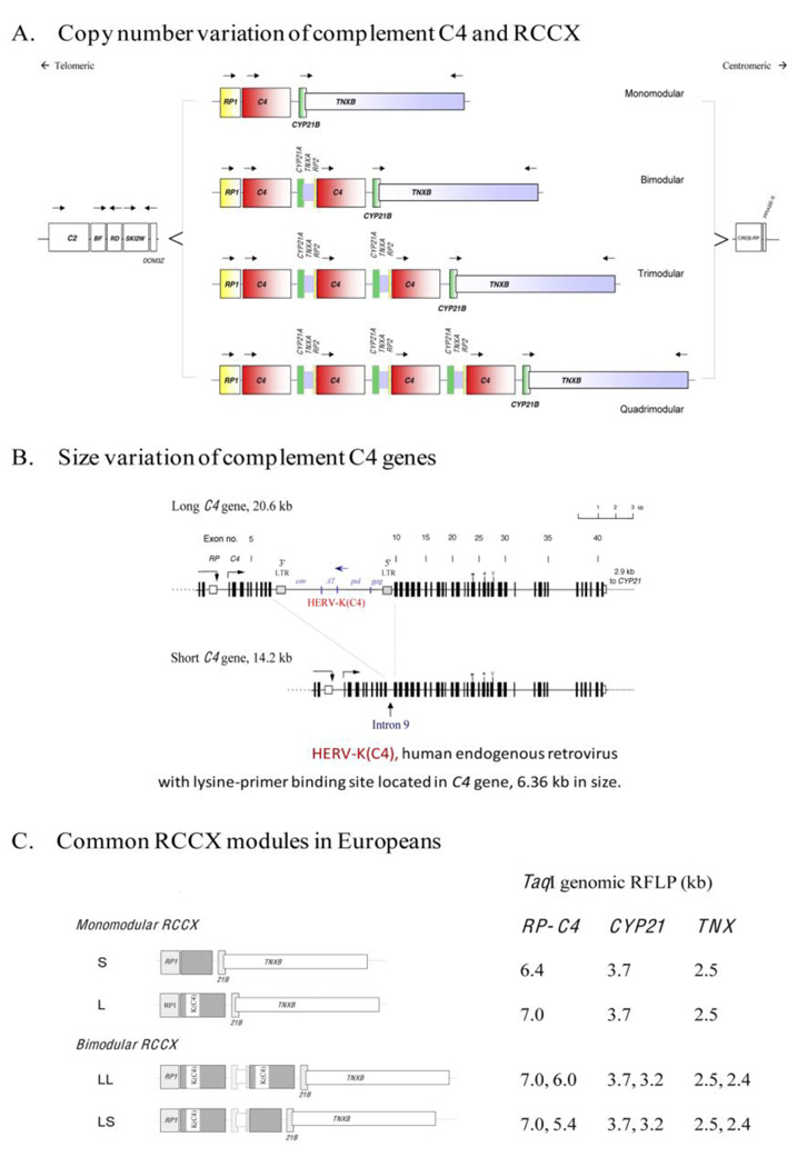
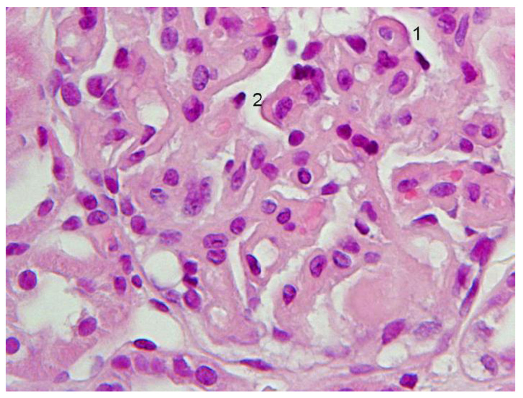
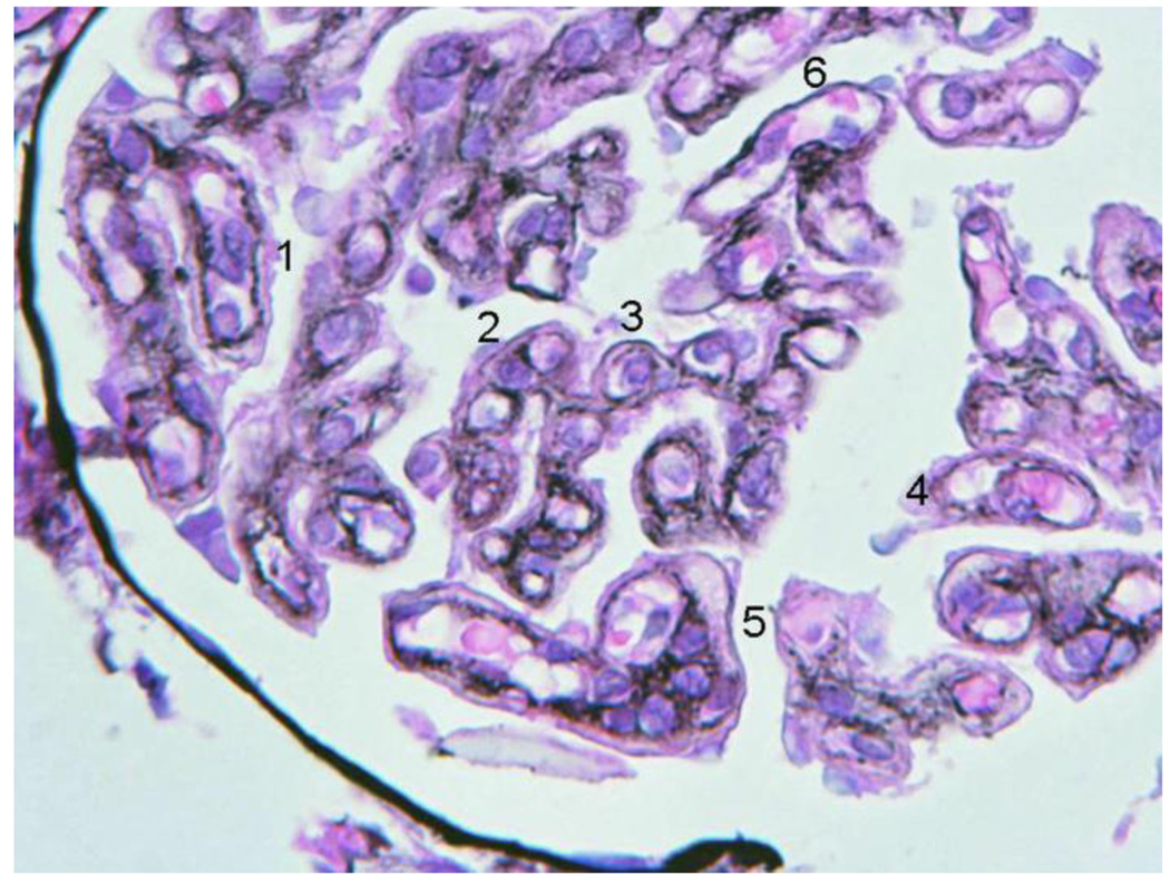
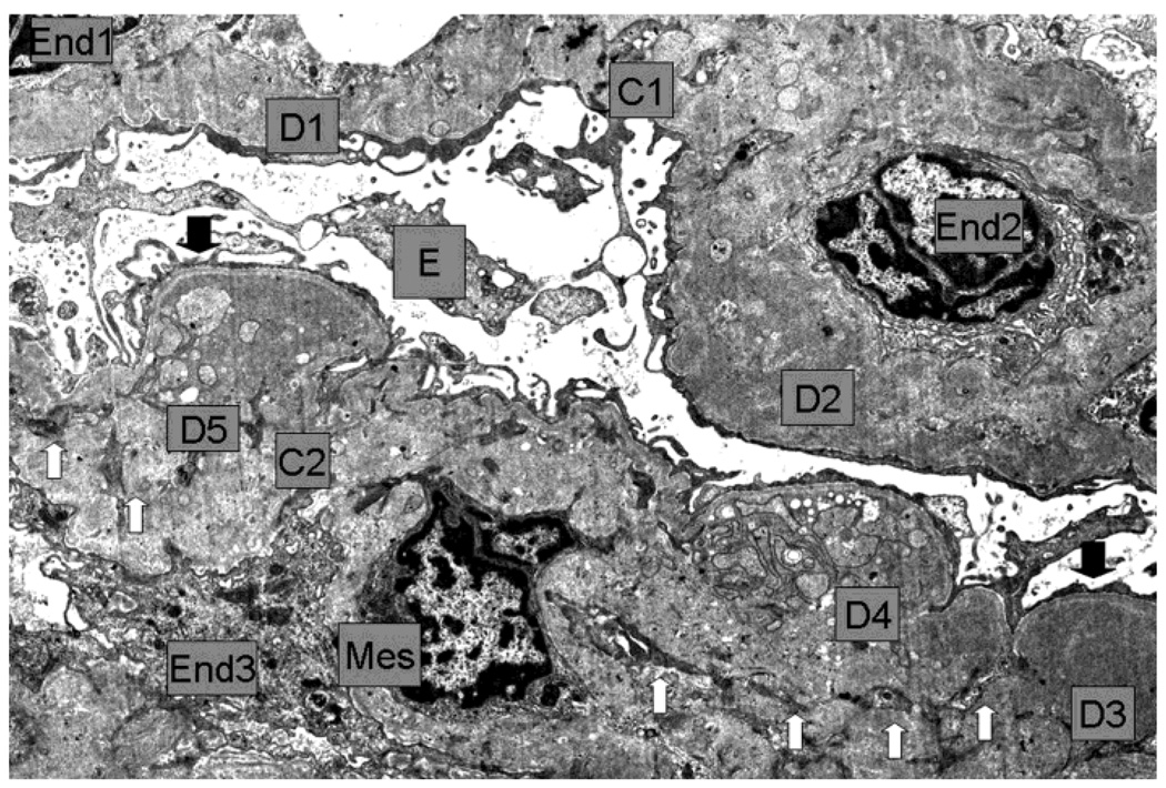
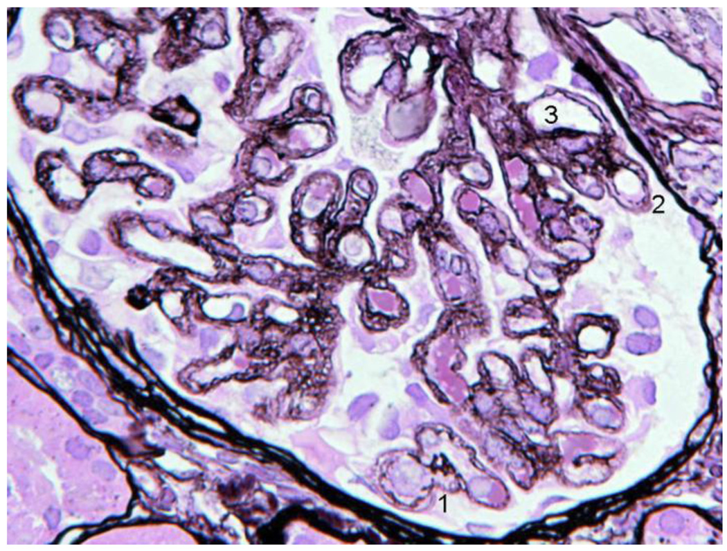
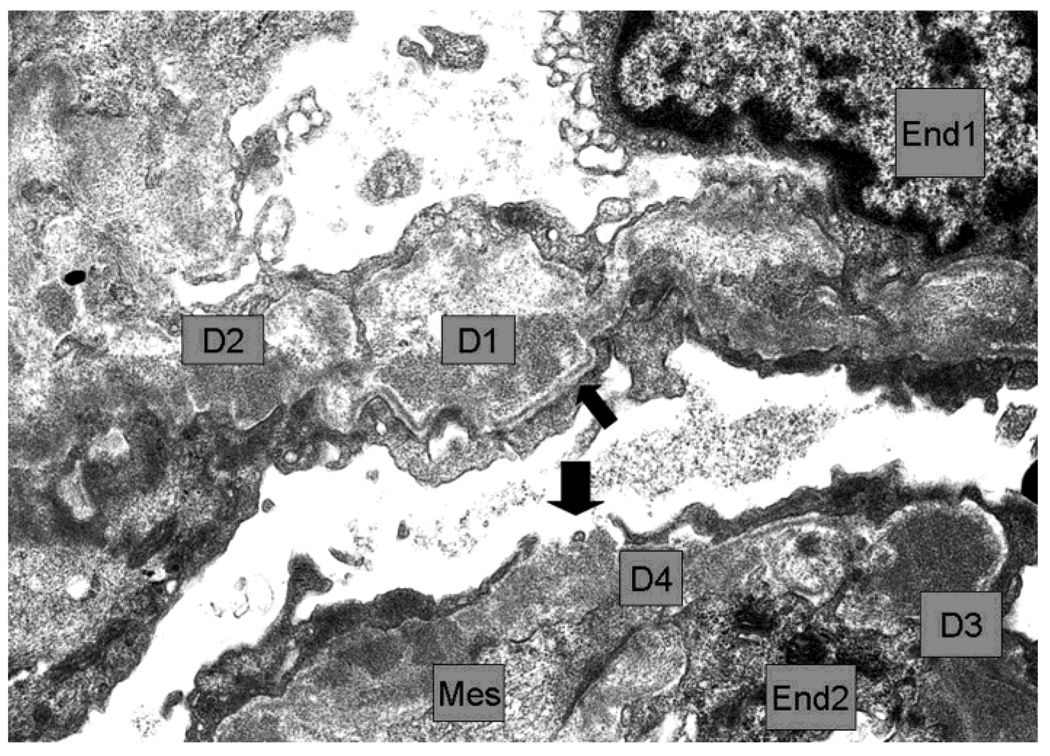
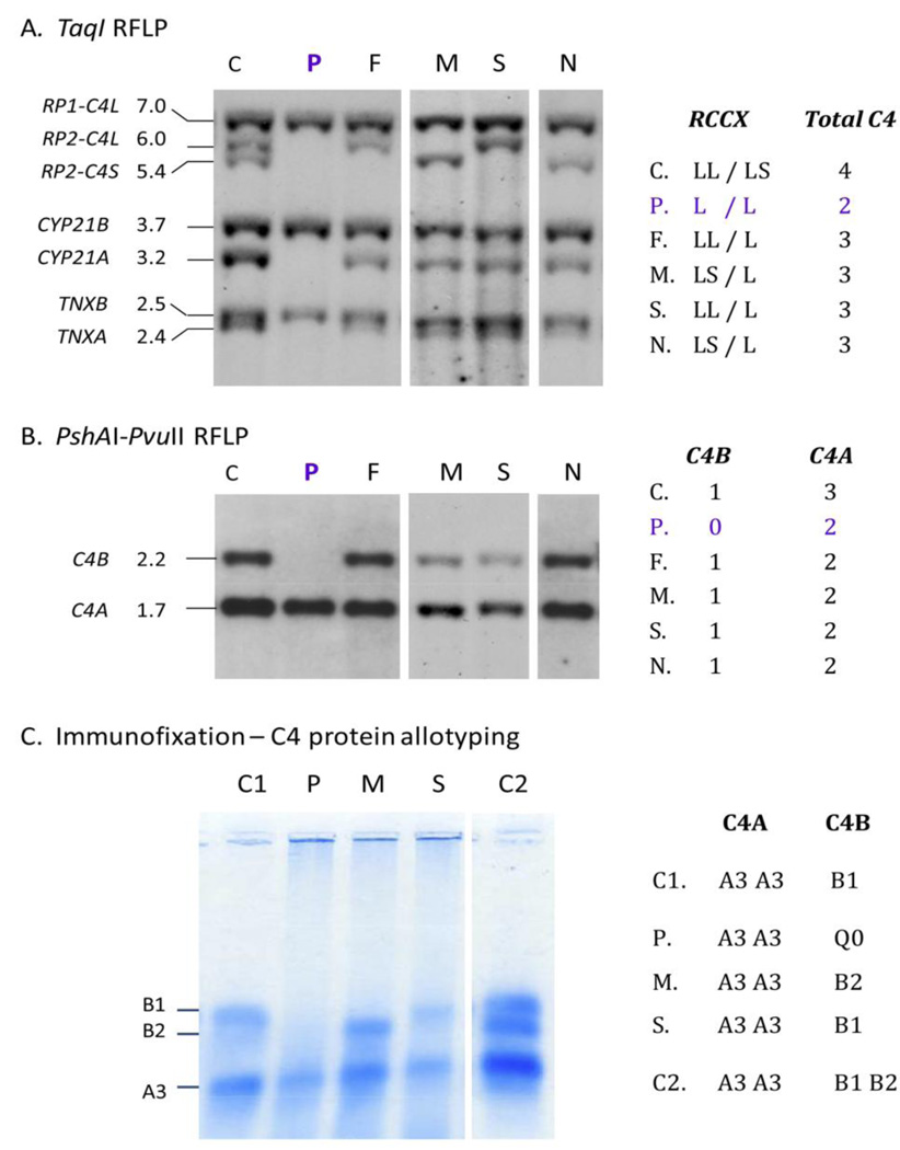
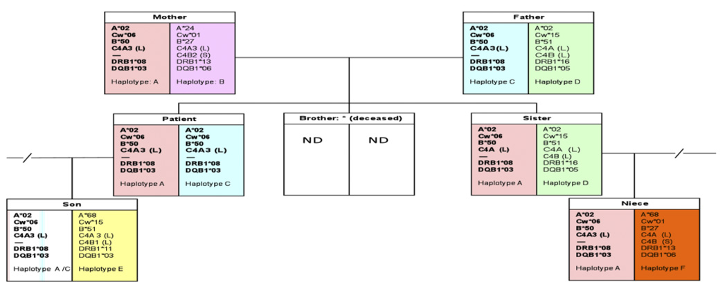
Homozygous complement C4B deficiency is described in a Southern European young female patient with Membranoproliferative Glomerulonephritis (MPGN) type III characterized by renal biopsies with strong complement C4 and IgG deposits. Low C4 levels were independent of clinical evolution or type of immunosuppression and were found in three other family members without renal disease or infections. HLA typing revealed that the patient has homozygous A*02, Cw*06, B*50 at the class I region, and DRB1*08 and DQB1*03 at the class II region. Genotypic and phenotypic studies demonstrated that the patient has homozygous monomodular RCCX in the HLA class III region, with single long C4A genes coding for C4A3 and complete C4B deficiency. Her father, mother, son and niece have heterozygous C4B deficiency. The patient's deceased brother had a history of Henoch-Schönlein Purpura (HSP), an immune complex-mediated proliferative glomerulonephritis. These findings challenge the putative pathophysiological roles of C4A and C4B and underscore the need to perform functional assays, C4 allotyping and genotyping on patients with persistently low serum levels of a classical pathway complement component and glomerulopathy associated with immune deposits.


Conflict of interest statement

Conflict of interest statement. None

Figures

Figure 1
Figure 1
The gene size and polygenic variations of human complement C4 and RP-C4-CYP21-TNX (RCCX modules) in the MHC class III region. A. Copy number variation of RCCX modules; B. Exon-intron structure and size variation of C4 gene; HERV-K(C4) is an endogenous virus inserted into intron 9 of long C4 genes; C. Common RCCX haplotypes and diagnostic TaqI restriction fragment size (in kb) for each haplotype (modified from ref. [50]).
Figure 2
Figure 2
First renal biopsy of the patient: Light microscopy (A) Silver stain (B), immunofluorescence (C), electromicroscopy (D) 2A Detail of the diffuse glomerulonephritis showing prominent generalized thickening of the capillary walls, sometimes with a broad glassy eosinophilic appearance (1, 2). There was slight to moderate increase in mesangial cells and expansion of the mesangial regions. Capillary lumina appear generally reduced. H+E. 2B. Within the thickened capillary walls the basement membrane appears irregular, interrupted or vacuolated (1, 3, 4). There are also several double contours some of which with a fairly thin outer contour (2, 3, 5). Any existing deposits within the wall appeared unstained with silver (5). One capillary wall has relatively normal wall (6). Mesangial matrix was mildly increased (5). Jones´s silver. 2C Intense confluent granular deposits of C4 with pseudolinear pattern globally distributed within the capillary walls and less intense mesangial staining. Large intense globular or semilunar collections in the wall were also seen. 2D Low magnification E.M: Capillaries (C1, C2) with endothelium (End1 to End3), Bowman´s space and visceral epithelium (E). In the thickened and deformed wall there are large, numerous moderately dense deposits (D1 to D5) more concentrated towards the subepithelial zone, but also extending through the whole wall (D2). Generally the dense deposits have uniform texture (D1 to D3) but some show complex irregular texture which includes less dense vesicular profiles, small clear spaces and groups of convoluted fine plasma membrane like structures (D4, D5). Generally there was no definition of the normal lamina densa of the basement membrane. Reactive type continuous thin lamina densa was seen along the epithelial side effectively separating the dense deposits from the podocytes (between black arrows). Clear cut formation of spikes was not seen. Reactive thin lamina densa on the endothelial side was ill defined and discontinuous. Mesangial cell (Mes) interposition was represented by multiple small cytoplasmic processes (white arrows) within the thickened capillary wall. There was severe thinning of podocytes and extensive effacement of foot processes. Negative magnification 2500×.
Figure 2
Figure 2
First renal biopsy of the patient: Light microscopy (A) Silver stain (B), immunofluorescence (C), electromicroscopy (D) 2A Detail of the diffuse glomerulonephritis showing prominent generalized thickening of the capillary walls, sometimes with a broad glassy eosinophilic appearance (1, 2). There was slight to moderate increase in mesangial cells and expansion of the mesangial regions. Capillary lumina appear generally reduced. H+E. 2B. Within the thickened capillary walls the basement membrane appears irregular, interrupted or vacuolated (1, 3, 4). There are also several double contours some of which with a fairly thin outer contour (2, 3, 5). Any existing deposits within the wall appeared unstained with silver (5). One capillary wall has relatively normal wall (6). Mesangial matrix was mildly increased (5). Jones´s silver. 2C Intense confluent granular deposits of C4 with pseudolinear pattern globally distributed within the capillary walls and less intense mesangial staining. Large intense globular or semilunar collections in the wall were also seen. 2D Low magnification E.M: Capillaries (C1, C2) with endothelium (End1 to End3), Bowman´s space and visceral epithelium (E). In the thickened and deformed wall there are large, numerous moderately dense deposits (D1 to D5) more concentrated towards the subepithelial zone, but also extending through the whole wall (D2). Generally the dense deposits have uniform texture (D1 to D3) but some show complex irregular texture which includes less dense vesicular profiles, small clear spaces and groups of convoluted fine plasma membrane like structures (D4, D5). Generally there was no definition of the normal lamina densa of the basement membrane. Reactive type continuous thin lamina densa was seen along the epithelial side effectively separating the dense deposits from the podocytes (between black arrows). Clear cut formation of spikes was not seen. Reactive thin lamina densa on the endothelial side was ill defined and discontinuous. Mesangial cell (Mes) interposition was represented by multiple small cytoplasmic processes (white arrows) within the thickened capillary wall. There was severe thinning of podocytes and extensive effacement of foot processes. Negative magnification 2500×.
Figure 3
Figure 3
Time course of proteinuria in response to different therapeutic approaches CF: Cyclophosphamide, 6 1 g monthly pulses PRED: prednisolone initially 1 mg/kg four month and then tapering MMF: Mycophenolate Mophetil 0,5-1g twice-a-day ACEI: angiotensin converting enzyme inhibitors RPC: ratio of urinary protein/creatinine (g/g) Alb: serum albumin (g/dL) C4 - Complement component C4 mg/dL (normal range 17.4–52.2)
Figure 4
Figure 4
Second Biopsy: silver stain LM (A), EM (B) 4A. The basement membrane was generally irregular, sometimes interrupted (1,2) and double contours were present (3). The vacuolated or moth eaten profile of the walls, seen in the first biopsy, was widespread and also present in the paramesangium. Capillaries with normal single layer basement membrane were not seen. The glomerular capsule appeared marginally thicker than in the first biopsy. 4B. The walls of 2 capillaries with endothelial cells (End1 and End2) contained several dense deposits (examples D1 to D4). D1 appeared rarified and surrounded by thin reactive basement membrane which appeared well-defined and continuous in the subepithelium (thin arrow) and discontinuous in the subendothelium. D2 made direct contact with the podocyte. D3 had fairly uniform high density and was also partly surrounded by subepithelial basement membrane. There was a gap between podocyte processes leaving D4 in direct contact with Bowman´s space. Mesangial cell cytoplasm (Mes) interposition was seen in the wall near D4 and other deposits. Negative 6000×
Figure 4
Figure 4
Second Biopsy: silver stain LM (A), EM (B) 4A. The basement membrane was generally irregular, sometimes interrupted (1,2) and double contours were present (3). The vacuolated or moth eaten profile of the walls, seen in the first biopsy, was widespread and also present in the paramesangium. Capillaries with normal single layer basement membrane were not seen. The glomerular capsule appeared marginally thicker than in the first biopsy. 4B. The walls of 2 capillaries with endothelial cells (End1 and End2) contained several dense deposits (examples D1 to D4). D1 appeared rarified and surrounded by thin reactive basement membrane which appeared well-defined and continuous in the subepithelium (thin arrow) and discontinuous in the subendothelium. D2 made direct contact with the podocyte. D3 had fairly uniform high density and was also partly surrounded by subepithelial basement membrane. There was a gap between podocyte processes leaving D4 in direct contact with Bowman´s space. Mesangial cell cytoplasm (Mes) interposition was seen in the wall near D4 and other deposits. Negative 6000×
Figure 5
Figure 5
Genotyping and phenotyping of complement C4. A. TaqI restriction fragment length polymorphism (RFLP) and Southern blot analyses of RCCX modules. B. PshAI-PvuII RFLP for C4A and C4B. C. Immunofixation of C4A and C4B proteins. Abbreviations: P, index patient; F, father; M, mother, S, son; N, niece; C, C1 and C2, three different healthy control subjects.
Figure 6
Figure 6
Family tree with HLA and complement C4 genotypes. One of the patient’s siblings was deceased and no genetic data was available.

Similar articles

Cited by

References

    1. Walport MJ. Complement-part I. New Engl. J. Med. 2001;344:1058–1066. - PubMed
    1. Chung EK, Wu YL, Yang Y, Zhou B, Yu CY. Human complement components C4A and C4B genetic diversities: complex genotypes and phenotypes. Curr. Protoc. Immunol. 2005:13.8.1–13.8.36. - PubMed
    1. Blanchong CA, Zhou B, Rupert KL, Chung EK, Jones KN, Sotos JF, Rennebohm RM, Yu CY. Deficiencies of human complement component C4A and C4B and heterozygosity in length variants of RP-C4-CYP21-TNX (RCCX) modules in Caucasians: the load of RCCX genetic diversity on MHC-associated disease. J. Exp. Med. 2000;191:2183–2196. - PMC - PubMed
    1. Yang Y, Chung EK, Wu YL, Savelli SL, Nagaraja HN, Zhou B, Hebert M, Jones KN, Shu Y, Kitzmiller K, Blanchong CA, McBride K, Higgins GC, Rennebohm RM, Rice RR, Hackshaw KV, Roubey RA, Grossman JM, Tsao BP, Birmingham DJ, Rovin BH, Hebert LA, Yu CY. Gene copy number variation and associated polymorphisms of complement component C4 in human systemic erythematosus (SLE): low copy number is a risk factor for and high copy number is a protective factor against European American SLE disease susceptibility. Am. J. Hum. Genet. 2007;80:1037–1054. - PMC - PubMed
    1. Yang Y, Chung EK, Zhou B, Lhotta K, Hebert LA, Birmingham DJ, Rovin BH, Yu CY. The intricate role of complement C4 in human systemic lupus erythematosus. Curr. Direct. Autoimmun. 2004;7:98–132. - PubMed

Publication types

MeSH terms