

Myoblast fusion in Drosophila
- PMID: 20580706
- PMCID: PMC2992827
- DOI: 10.1016/j.yexcr.2010.05.018
Myoblast fusion in Drosophila
Abstract
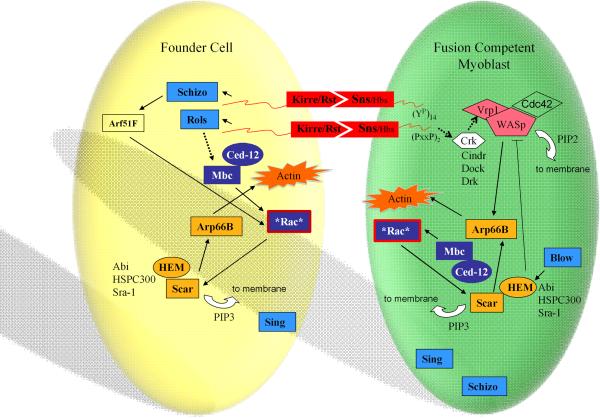
The body wall musculature of a Drosophila larva is composed of an intricate pattern of 30 segmentally repeated muscle fibers in each abdominal hemisegment. Each muscle fiber has unique spatial and behavioral characteristics that include its location, orientation, epidermal attachment, size and pattern of innervation. Many, if not all, of these properties are dictated by founder cells, which determine the muscle pattern and seed the fusion process. Myofibers are then derived from fusion between a specific founder cell and several fusion competent myoblasts (FCMs) fusing with as few as 3-5 FCMs in the small muscles on the most ventral side of the embryo and as many as 30 FCMs in the larger muscles on the dorsal side of the embryo. The focus of the present review is the formation of the larval muscles in the developing embryo, summarizing the major issues and players in this process. We have attempted to emphasize experimentally-validated details of the mechanism of myoblast fusion and distinguish these from the theoretically possible details that have not yet been confirmed experimentally. We also direct the interested reader to other recent reviews that discuss myoblast fusion in Drosophila, each with their own perspective on the process [1-4]. With apologies, we use gene nomenclature as specified by Flybase (http://flybase.org) but provide Table 1 with alternative names and references.
Copyright © 2010 Elsevier Inc. All rights reserved.
Figures
Similar articles
-
Expression and functional analysis of a novel Fusion Competent Myoblast specific GAL4 driver.Gene Expr Patterns. 2008 Jan;8(2):87-91. doi: 10.1016/j.modgep.2007.10.002. Epub 2007 Oct 13. Gene Expr Patterns. 2008. PMID: 17988956 Free PMC article.
-
3D analysis of founder cell and fusion competent myoblast arrangements outlines a new model of myoblast fusion.Dev Biol. 2007 Sep 1;309(1):113-25. doi: 10.1016/j.ydbio.2007.06.024. Epub 2007 Jul 6. Dev Biol. 2007. PMID: 17662708 Free PMC article.
-
Variation in mesoderm specification across Drosophilids is compensated by different rates of myoblast fusion during body wall musculature development.PLoS One. 2011;6(12):e28970. doi: 10.1371/journal.pone.0028970. Epub 2011 Dec 14. PLoS One. 2011. PMID: 22194964 Free PMC article.
-
Cell and molecular biology of myoblast fusion.Int Rev Cytol. 2003;225:33-89. doi: 10.1016/s0074-7696(05)25002-7. Int Rev Cytol. 2003. PMID: 12696590 Review.
-
Myoblast fusion in Drosophila.Bioessays. 2002 Jul;24(7):591-601. doi: 10.1002/bies.10115. Bioessays. 2002. PMID: 12111720 Review.
Cited by
-
Characterization of early steps in muscle morphogenesis in a Drosophila primary culture system.Fly (Austin). 2011 Apr-Jun;5(2):68-75. doi: 10.4161/fly.5.2.15031. Epub 2011 Apr 1. Fly (Austin). 2011. PMID: 21339707 Free PMC article.
-
Ferlin proteins in myoblast fusion and muscle growth.Curr Top Dev Biol. 2011;96:203-30. doi: 10.1016/B978-0-12-385940-2.00008-5. Curr Top Dev Biol. 2011. PMID: 21621072 Free PMC article. Review.
-
Generation of a monoclonal antibody reactive to prefusion myocytes.J Muscle Res Cell Motil. 2011 Aug;32(1):31-8. doi: 10.1007/s10974-011-9247-8. Epub 2011 May 20. J Muscle Res Cell Motil. 2011. PMID: 21597958 Free PMC article.
-
The Formin Diaphanous Regulates Myoblast Fusion through Actin Polymerization and Arp2/3 Regulation.PLoS Genet. 2015 Aug 21;11(8):e1005381. doi: 10.1371/journal.pgen.1005381. eCollection 2015 Aug. PLoS Genet. 2015. PMID: 26295716 Free PMC article.
-
Spatiotemporal regulation of the GPCR activity of BAI3 by C1qL4 and Stabilin-2 controls myoblast fusion.Nat Commun. 2018 Oct 26;9(1):4470. doi: 10.1038/s41467-018-06897-5. Nat Commun. 2018. PMID: 30367035 Free PMC article.
References
-
- Onel SF, Renkawitz-Pohl R. FuRMAS: triggering myoblast fusion in Drosophila. Dev. Dyn. 2009;238:1513–1525. - PubMed
-
- Abmayr SM, Zhuang S, Geisbrecht ER. Myoblast fusion in Drosophila. Methods Mol. Biol. 2008;475:75–97. - PubMed
-
- Abmayr SM, Balagopalan L, Galletta BJ, Hong SJ, Lawrence IG, Kostas I, Sarjeet SG. Comprehensive Molecular Insect Science. Elsevier; Amsterdam: 2005. Myogenesis and Muscle Development; pp. 1–43.
Publication types
MeSH terms
Grants and funding
LinkOut - more resources
Full Text Sources
Molecular Biology Databases
