. 2010 Sep;139(3):869-81, 881.e1-9.
doi: 10.1053/j.gastro.2010.05.037. Epub 2010 May 24.

Phosphoinositide 3-kinase signaling mediates beta-catenin activation in intestinal epithelial stem and progenitor cells in colitis

Affiliations

Phosphoinositide 3-kinase signaling mediates beta-catenin activation in intestinal epithelial stem and progenitor cells in colitis

Goo Lee et al. Gastroenterology. 2010 Sep.

Abstract

Background & aims: Mechanisms responsible for crypt architectural distortion in chronic ulcerative colitis (CUC) are not well understood. Data indicate that serine/threonine protein kinase Akt (Akt) signaling cooperates with Wingless (Wnt) to activate beta-catenin in intestinal stem and progenitor cells through phosphorylation at Ser552 (P-beta-catenin(552)). We investigated whether phosphoinositide 3-kinase (PI3K) is required for Akt-mediated activation of beta-catenin during intestinal inflammation.

Methods: The class IA subunit of PI3K was conditionally deleted from intestinal epithelial cells in mice named I-pik3r1KO. Acute inflammation was induced in mice and intestines were analyzed by biochemical and histologic methods. The effects of chemically blocking PI3K in colitic interleukin-10(-/-) mice were examined. Biopsy samples from patients were examined.

Results: Compared with wild-type, I-pik3r1KO mice had reduced T-cell-mediated Akt and beta-catenin signaling in intestinal stem and progenitor cells and limited crypt epithelial proliferation. Biochemical analyses indicated that PI3K-Akt signaling increased nuclear total beta-catenin and P-beta-catenin(552) levels and reduced N-terminal beta-catenin phosphorylation, which is associated with degradation. PI3K inhibition in interleukin-10(-/-) mice impaired colitis-induced epithelial Akt and beta-catenin activation, reduced progenitor cell expansion, and prevented dysplasia. Human samples had increased numbers of progenitor cells with P-beta-catenin(552) throughout expanded crypts and increased messenger RNA expression of beta-catenin target genes in CUC, colitis-associated cancer, tubular adenomas, and sporadic colorectal cancer, compared with control samples.

Conclusions: PI3K-Akt signaling cooperates with Wnt to increase beta-catenin signaling during inflammation. PI3K-induced and Akt-mediated beta-catenin signaling are required for progenitor cell activation during the progression from CUC to CAC; these factors might be used as biomarkers of dysplastic transformation in the colon.


Figures

Figure 1
Figure 1. Loss of p85α protein expression in IECs of I-pik3r1 KO mice
Western blot analyses of total p85, p85α and p110α subunits of PI3K in cytosolic fractions from purified crypt epithelial cells at 0 and 3 hours after T cell activation with anti-CD3 mAb in pik3r1lox/lox and I-pik3r1 KO mice. h, hours.
Figure 2
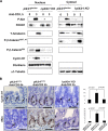
Figure 2. Epithelial deletion of pik3r1 attenuates T cell-mediated Akt and β-catenin signaling in SB crypts
(A) Western blot analyses of P-Akt, FoxO1, total (T) β-catenin, P-β-catenin/,, P-β-catenin552, and cyclin D1 in nuclear or cytoplasmic extracts of purified SB crypt epithelial cells from pik3r1lox/lox and I-pik3r1 KO mice following T cell activation. Fibrillarin and α1-Tubulin are subcellular fraction controls. (B) Cyclin D1 and cMyc in SB crypts detected by immunohistochemistry (arrows). Bar graphs show mean ± standard deviation (±SD) of positive cells/crypt in indicated groups. Scale bar size is 50μm. p-value represents the results of student t-test.
Figure 3
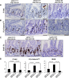
Figure 3. PI3K is required for T cell mediated Akt and β-catenin signaling in SB crypts
Immunohistochemistry staining for (A) P-Akt, (B) P-β-catenin552 (arrows) performed in ileal sections of control and anti-CD3 treated pik3r1lox/lox or I-pik3r1 KO mice. Insets indicate P-Akt and P-β-catenin552 positive epithelial cells with enlarged nuclei within the crypt. (C) BrdU incorporation was examined 18 hrs after anti-CD3 treatment. (D) Bar graphs display mean ±SD of positively stained cells/crypt for P-Akt, P-β-catenin552, and BrdU.
Figure 4
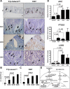
Figure 4. PI3K is required for epithelial cell proliferation induced by T cell activation
(A) Single crypt picture indicating enumeration of crypt cells. (B) P-β-catenin552 and (C) BrdU labeled cells per position of SB crypt determined 3 or 18 hours after T cell activation, respectively. (D) TUNEL staining of control pik3r1lox/lox or pik3r1lox/lox and I-pik3r1 KO mice 18hr after anti-CD3 mAb injection. (E) Bar graph quantifying D shows mean ±SD of TUNEL-positive cells/crypt. Scale bar size is 50μm. p-value represents results of student t- test. (F) Representative image and quantification of a P-β-catenin552 positive CBC (block arrow) with 2 neighboring paneth cells (small arrows) in the SB. 100 well-oriented SB crypts were counted 3hrs after control or anti-CD3 treatment.
Figure 5
Figure 5. PI3K is required for progression of chronic CID
(A) Timeline of colitis induction with Px in IL10−/− mice and LY294002 treatment. (B) Gross findings in colons of WT, or vehicle only and LY294002 D42 treated IL10−/− mice. (C) Ulcerating masses (red arrows) in methylene blue-stained colonic mucosa. (D) Prevalence of dysplasia in indicated group over time. (E) Western blot data showed cytosolic P-Akt and nuclear P-β-catenin in IECs. Bar size=500μm.
Figure 6
Figure 6. Immunohistochemical evidence that PI3K is required for progression of CID
(A) Immunohistochemistry for P-Akt, P-β-catenin552, cyclin D1, or BrdU incorporation in colonic sections of control, D42-colitis, D42-dysplasia, D42-LY294002-treated IL10−/− mice. (B) Bar graph shows mean ±SD of positively stained cells of P-Akt, P-β-catenin552, cyclin D1, and BrdU in each group. *p<0.01, compared to control; **p<0.01, compared to D42-colitis, #p<0.01, compared to D42-dysplasia. Bar size=50μm. p-value represents results of student t-test.
Figure 7
Figure 7. β-catenin activation and Ki67 levels are increased in CUC, UCD, and CAC patients
(A) Human colonic specimens stained for P-β-catenin552 and Ki67. Arrows indicate positively stained cells in representative control (C), active non-treated CUC, UCD, and CAC samples. Block arrow shows P-β-catenin552 stained cells at points of crypt branching (dotted line). Scale bar size=50μm. (B) Bar graphs show mean ±SD of P-β-catenin552- and (C) Ki67-stained cells/100 epithelial cells in each group. The number of patients in each group in B is 20 C, 13 CUC, 10 UCD, and 2 CAC, and in C is 8 C, 10 CUC, 4 UCD, and 2 CAC. (D) Real-time RT-PCR for β-catenin target genes in human colonic tissues. Bar graphs show mean ±SD of mRNA fold-induction of cMyc (Myc), COX2 (PTGS2), and GPR49 (LGR5) in the indicated groups. Data represents specimens from 11–13 C, 19–24 CUC, 4–5 UCD, and 5–7 CAC patients. p-value represents the results of a Mann-Whitney U test. (E) Wnt signaling inhibits the destruction complex, impairs N-terminal phoshorylation (degradation), and stabilizes β-catenin which accumulates in the nucleus. In the inflammatory setting PI3K/Akt signaling is activated and β-catenin is phosphorylated at Ser552. The location for this phosphorylaton event is unknown, but P-β-catenin552 is only detected in the nucleus in mice. Nuclear P-β-catenin552 binds to the Tcf4 to induce transcription.

Comment in

References

    1. Franklin WA, McDonald GB, Stein HO, et al. Immunohistologic demonstration of abnormal colonic crypt cell kinetics in ulcerative colitis. Hum Pathol. 1985;16:1129–32. - PubMed
    1. Jenkins D, Balsitis M, Gallivan S, et al. Guidelines for the initial biopsy diagnosis of suspected chronic idiopathic inflammatory bowel disease. The British Society of Gastroenterology Initiative. J Clin Pathol. 1997;50:93–105. - PMC - PubMed
    1. Cheng H, Bjerknes M, Amar J, Gardiner G. Crypt production in normal and diseased human colonic epithelium. Anat Rec. 1986;216:44–8. - PubMed
    1. Bjerknes M, Cheng H. Gastrointestinal stem cells. II. Intestinal stem cells. Am J Physiol Gastrointest Liver Physiol. 2005;289:G381–7. - PubMed
    1. Gregorieff A, Pinto D, Begthel H, et al. Expression pattern of Wnt signaling components in the adult intestine. Gastroenterology. 2005;129:626–38. - PubMed

Publication types

MeSH terms