. 2010 Oct;40(1):200-6.
doi: 10.1016/j.nbd.2010.05.025. Epub 2010 May 23.

Alcohol-induced neuroapoptosis in the fetal macaque brain

Affiliations

Alcohol-induced neuroapoptosis in the fetal macaque brain

Nuri B Farber et al. Neurobiol Dis. 2010 Oct.

Abstract

The ability of brief exposure to alcohol to cause widespread neuroapoptosis in the developing rodent brain and subsequent long-term neurocognitive deficits has been proposed as a mechanism underlying the neurobehavioral deficits seen in fetal alcohol spectrum disorder (FASD). It is unknown whether brief exposure to alcohol causes apoptosis in the fetal primate brain. Pregnant fascicularis macaques at various stages of gestation (G105 to G155) were exposed to alcohol for 8h, then the fetuses were delivered by caesarean section and their brains perfused with fixative and evaluated for apoptosis. Compared to saline control brains, the ethanol-exposed brains displayed a pattern of neuroapoptosis that was widespread and similar to that caused by alcohol in infant rodent brain. The observed increase in apoptosis was on the order of 60-fold. We propose that the apoptogenic action of alcohol could explain many of the neuropathological changes and long-term neuropsychiatric disturbances associated with human FASD.


Figures

Figure 1
Figure 1
An image from the somatosensory cortex of a G155 NHP fetus exposed in utero to alcohol. Panel A was stained with AC3 antibodies and Panel B is an adjacent section stained by the DeOlmos cupric silver method. Note that each stain detects the same population of neurons, which are distributed primarily in a single superficial layer of the neocortex. Other neuronal populations throughout the brain that were selectively AC3-positive were also selectively silver positive. Mag: 130x.
Figure 2
Figure 2
Anterior cingulate cortex of NHP fetuses exposed to alcohol (Panels A & B) or saline (Panel C) at G120 (Panel A) or G155 (Panels B & C) and stained by the de Olmos cupric silver method. Following alcohol exposure at G120 (Panel A), apoptotic neurons are concentrated in deeper layers, whereas after alcohol at G155 (Panel B), apoptotic neurons are concentrated more superficially. Controls exposed to saline at either age had rare or no apoptotic neurons in either layer (Panel C). Mag: Panel A = 150x; Panels B & C = 130x.
Figure 3
Figure 3
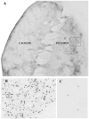
Panel A is a section through the caudate-putamen from a G120 fetal NHP brain following alcohol exposure showing a dense pattern of neuroapoptosis (AC3-positive staining) in both regions, especially putamen. Each tiny dark speck is a degenerating neuron. Panel B is from the boxed region in A showing apoptotic putamen neurons at higher magnification. Panel C is from the putamen of a G120 control brain demonstrating that the physiological cell death rate under normal circumstances is very low. Caudate-putamen neurons respond very early to alcohol and by 8 hrs the stained cells have a shrunken and irregular shape reflecting deteriorative changes. The pattern of degeneration in the NHP caudate-putamen following alcohol treatment is remarkedly similar to that which occurs in the infant mouse caudate-putamen following alcohol treatment (Olney et al., 2002b, Figure 3). Mag: Panel A = 10x; Panels B & C = 55x.
Figure 4
Figure 4
AC3-stained section from the subiculum of the fetal NHP brain in early gestation (G120 following exposure for 8 hrs to saline or alcohol. In the saline-treated brain there are very few AC3-positive neuronal profiles (none in this scene), whereas in the alcohol-treated brain such profiles are abundant. We judge these cells to be in an early stage of degeneration based on their display of a relatively intact and normal-appearing microanatomy. This pattern of degeneration in the NHP subiculum shows a strong resemblance to that we have demonstrated (Olney et al., 2002, Figure 3) in the infant mouse subiculum following alcohol treatment. Mag 75x.
Figure 5
Figure 5
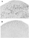
AC3-stained sections from the inferior colliculus of the NHP fetal brain at G130-135 following alcohol (Panel A) or saline (Panel B). Similar to what is seen in the superior colliculus (Tenkova et al., 2003), inferior collicular neurons are relatively more sensitive than most neuronal populations and begin degenerating early. Therefore, by 8 hrs immunoreactivity is receding and is primarily confined to the cell body. There is an abundance of AC3-positive profiles in the alcohol-exposed inferior colliculus and very few such profiles following saline. Mag 100x.
Figure 6
Figure 6
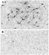
AC3-stained sections from the anterdorsal (AD) nucleus of the thalamus of the fetal NHP brain (gestation age 130-135 days) following alcohol (A) or saline (B). In the section from the saline-treated animal there is one or possibly two faintly positive profile(s), whereas in the section from the alcohol-treated animal there are approximately 35 intensely immunoreactive profiles. The dendritic processes of the degenerating thalamic neurons (Panel A) are intensely AC3-positive, signifying that these neurons are in a relatively early stage of degeneration. The particulate debris in the vicinity of the degenerating dendrites signifies that they are beginning to deteriorate and become fragmented. As we have demonstrated previously (Olney et al., 2002b), Figure 5), alcohol has a similar predilection for damaging the AD thalamic nucleus in the infant mouse brain. Mag 180x.
Figure 7
Figure 7
Alcohol-induced neurodegeneration in the fetal NHP cerebellum following alcohol exposure at G105-110 (panel A) or G120 (Panel B). Panel C is from a control animal. AC3 staining. PCL = Purkinje cell layer. IGL = internal granule layer. Mag 40x.
Figure 8
Figure 8
Density of AC3-positive profiles in 5 NHP fetal brains exposed to alcohol compared to 4 NHP control brains. The mean (± SEM) density for the alcohol brains (5330 ± 668.40 profiles per mm3) was 61.8-fold higher than in the control brains (86.25 ± 27.75 profiles per mm3), a difference significant at p = 0.0014

Similar articles

Cited by

References

    1. Adams RD, Victor M. Principles of Neurology. McGraw-Hill Inc.; New York: 1989.
    1. Aggleton JP, Brown MW. Episodic memory, amnesia, and the hippocampal-anterior thalamic axis. Behav Brain Sci. 1999;22:425–44. discussion 444-89. - PubMed
    1. Archibald SL, Fennema-Notestine C, Gamst A, Riley EP, Mattson SN, Jernigan TL. Brain dysmorphology in individuals with severe prenatal alcohol exposure. Dev Med Child Neurol. 2001;43:148–54. - PubMed
    1. Bailey BN, Delaney-Black V, Covington CY, Ager J, Janisse J, Hannigan JH, Sokol RJ. Prenatal exposure to binge drinking and cognitive and behavioral outcomes at age 7 years. Am J Obstet Gynecol. 2004;191:1037–43. - PubMed
    1. Bauer-Moffett C, Altman J. The effect of ethanol chronically administered to preweanling rats on cerebellar development: a morphological study. Brain Res. 1977;119:249–68. - PubMed

Publication types

MeSH terms