Review
. 2010 Oct 1;588(Pt 19):3639-55.
doi: 10.1113/jphysiol.2010.189605.

Discovery and rediscoveries of Golgi cells

Affiliations
Review

Discovery and rediscoveries of Golgi cells

Elisa Galliano et al. J Physiol. .

Abstract

When Camillo Golgi invented the black reaction in 1873 and first described the fine anatomical structure of the nervous system, he described a ‘big nerve cell’ that later took his name, the Golgi cell of cerebellum (‘Golgi’schen Zellen’, Gustaf Retzius, 1892). The Golgi cell was then proposed as the prototype of type-II interneurons, which form complex connections and exert their actions exclusively within the local network. Santiago Ramón y Cajal (who received the Nobel Prize with Golgi in 1906) proceeded to a detailed description of Golgi cell morphological characteristics, but functional insight remained very limited for many years. The first rediscovery happened in the 1960s, when neurophysiological analysis in vivo revealed that Golgi cells are inhibitory interneurons. This finding promoted the development of two major cerebellar theories, the ‘beam theory’ of John Eccles and the ‘motor learning theory’ of David Marr, in which the Golgi cells regulate the spatial organisation and the gain of input signals to be processed and learned by the cerebellar circuit. However, the matter was not set and a series of pioneering observations using single unit recordings and electronmicroscopy raised new issues that could not be fully explored until the 1990s. Then, the advent of new electrophysiological and imaging techniques in vitro and in vivo demonstrated the cellular and network activities of these neurons. Now we know that Golgi cells, through complex systems of chemical and electrical synapses, effectively control the spatio-temporal organisation of cerebellar responses. The Golgi cells regulate the timing and number of spikes emitted by granule cells and coordinate their coherent activity. Moreover, the Golgi cells regulate the induction of long-term synaptic plasticity along the mossy fibre pathway. Eventually, the Golgi cells transform the granular layer of cerebellum into an adaptable spatio-temporal filter capable of performing several kinds of logical operation. After more than a century, Golgi’s intuition that the Golgi cell had to generate under a new perspective complex ensemble effects at the network level has finally been demonstrated.


Figures

Figure 1
Figure 1
Camillo Golgi, the black reaction and the Golgi cell A, Camillo Golgi, Nobel Prize winner for Phsyiology and Medicine (1906). Camillo Golgi (1843–1926) invented the ‘black reaction’, which allowed him and many other scientists to visualise the fine structure of the nervous system. B, in this picture, Golgi shows a reconstruction of a Golgi cell. Note the precise description of the basal and apical dendrites and of the large axonal plexus, the hallmark of the Golgi cell. (Gangliar cell in the (neonatal) cat cerebellar cortex; Table XIV, Opera Omnia, Golgi, 1903.) ‘Such cells are part of the granular layer and, in the cat and rabbit, when the black reaction succeeds, can be seen in a considerable amount. … Most ramifications of the protoplasmatic prolongations (in black) reach the superior border of the molecular layer. … The nervous prolongation (in red), with repeated subdivisions becoming finer and finer, creates an extremely complicated interlacement of fibres. These, in the vertical plane, spread from one to the other border of the granular layer, and in the width of the granular layer mix up with the interlacements resulting from subdivisions of neighbouring cells of the same type (see Table XVII). This cell is one of the most remarkable specimens among those that are described as second type cells in the text. Regarding the cerebellum, such a cell should be to compared to the one represented in Table XV (a Purkinje cell), representing one of the most remarkable specimens of the first type of cells.’ Translated from Opera Omnia (Golgi, 1903).
Figure 3
Figure 3
The relationship between Golgi cells and granule cells Granule cells largely exceed in number the Golgi cells and are much smaller (Purkinje cells are distinctly shown in the background). Therefore a Golgi cell can innervate several granule cells lying within their axonal plexus, capturing another fundamental feature of the granular layer organisation. (Fragment of a rabbit cerebellar convolution (vertical section); Table XIX, Opera Omnia, Golgi, 1903). ‘This drawing was specifically made to illustrate the granular layer. … The so-called granule cells look like nervous cells with a globose shape, really small and equipped with 3, 4, 5 or even 6 prolongations, among which just one has the features of nervous prolongation (the nervous prolongation is just outlined, red thread). Prolongations, which it seems to be correct to name protoplasmatic, even if they appear slightly different from other gangliar cells’ prolongations, end up with a small granulous mass, towards which neighbouring granule cells’ prolongation often converge. In the region in which the granular layer merges into the molecular layer, two large cells are drawn. These are placed laterally and differ from Purkinje cells for the cell body shape, for the way of branching of their protoplasmatic prolongations and, overall, for the very different organisation of the nervous prolongation. … These two large cells are of the same type of the ones already illustrated in Tables XIV and XVII.’ Translated from Opera Omnia (Golgi, 1903).
Figure 4
Figure 4
Golgi cell activity in vivo A, the Golgi cell shows rhythmic background activity in vivo (from Holtzman et al. 2006a). B, peripheral sensory stimulation elicits bursts of activity (from Holtzman et al. 2006a). Each burst is usually composed of 2–3 spikes and is followed by a long-lasting inhibitory period (or silent pause in Vos et al. 1999). C, during locomotion, the Golgi cell is entrained into repetitive activity cycles, during which its frequency is modulated.
Figure 8
Figure 8
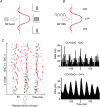
The physiological consequences of Golgi cell activation Golgi cell activation has complex consequences for granular layer responses. A, a burst in a group of mossy fibres (MF) generates stronger and faster granule cell responses in the centre than in the surround of a granular layer field (the red profile represents the excitatory/inhibitory balance in a granular layer field activated by a group of MFs and regulated by Golgi cells; drawn after Mapelli & D’Angelo, 2007). B, a MF theta-burst stimulation (TBS) in a group of MFs generates more effectively LTP in the centre and LTD in the surround of the granular layer field. C, the raster plot shows that a diffused random stimulation of the MFs generates a coherent response of the granule cells (GrC) and of the Golgi cells (GoC). Synchronisation is due to parallel fibre feed-back inhibition. Crosscorrelograms (CCH) are shown for two GoCs and for all the granule cells and the Golgi cells in the network revealing their coherence. Data elaborated from the model of Solinas et al. (2010).
Figure 6
Figure 6
Control of granular layer spatio-temporal dynamics by Golgi cell inhibition A, stimulation of a mossy fibre beam elicits local field potentials in the granular layer, which can be composed by more spikes generated in sequence by granule cells. Activation of the feed-forward inhibitory Golgi cell loop limits spike emission (time-window effect; from Mapelli & D’Angelo, 2007). B, stimulation of a mossy fibre beam elicits local field potentials in the granular layer, which are surrounded by lateral inhibition generated by Golgi cells (centre–surround effect; from Mapelli & D’Angelo, 2007).
Figure 2
Figure 2
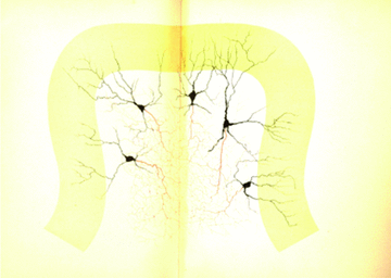
The spatial relationship between multiple Golgi cells Note that no restriction is imposed to axons, which spread and overlap into the granular layer, that apical dendrites ramify in the molecular layer without specific orientation, and that basal dendrites ramify in the granular layer over a surface smaller than that occupied by the axon. In this figure, the fundamental features of the Golgi cell are captured. (Fragment of a (neonatal) cat cerebellar convolution (vertical section); Table XVII, Opera Omnia, Golgi, 1903). ‘The drawing is specifically made to show shape, disposition, ramification laws, localisation and relationships of the large gangliar cells of the granular layer. Protoplasmatic prolongations branch dichotomously in a very different way compared to Pukinje cells. The most distal extensions of the branches often reach the molecular layer peripheral limit. Nervous prolongations, with their fine and repeated subdivisions, form a complicated interlacement with the result that it is impossible to follow the course of single prolongations. Such interlacement does not seem to have borders either toward the inside of the granular layer or toward the molecular layer. Thus, several of these interlacements obviously mix up to form a complicated plexus.’ Translated from Opera Omnia (Golgi, 1903).
Figure 7
Figure 7
The electrical activity of Golgi cells The Golgi cell shows response dynamics, which can support the cycles of activation and inactivation observed in various functional conditions (see Figs 5 and 6). A, the Golgi cell in vitro shows (1) pacemaker activity at around 7 Hz. In response to depolarisation (2), the Golgi cell shows high-frequency discharge with frequency adaptation. In response to hyperpolarisation (3), the Golgi cell shows sagging inward rectification followed by (4) rebound excitation. Bursts of activity are followed by (5) a silent pause. B, demonstration of the silent pause following a burst response to a mossy fibre stimulus (arrows in the inset). C, when stimulated with pulses repeated at different frequencies, the Golgi cell shows enhanced responses (faster and higher frequency spikes) at the resonant frequency of 6 Hz. The tracings are simulated from the models of Solinas et al. (2007a,, and reproduce response behaviours reported in Dieudonné (1988), Forti et al. (2006) and Solinas et al. (2007a,;.
Figure 5
Figure 5
Golgi cell network entrainment A, the Golgi cell spikes are in phase with the local field potential of the granular layer (Duguéet al. 2009). B, Golgi cells can show rhythmic entrainment with the UP–DOWN states characterising neocortical activity (Ros et al. 2009). The different behaviour in A and B may reflect different functional states or simply the fact that the trace in A may be part of an UP state as shown in B.

Similar articles

Cited by

References

    1. Albus JS. A theory of cerebellar function. Math Biosci. 1971;10:25–61.
    1. Ambrosi G, Flace P, Lorusso L, Girolamo F, Rizzi A, Bosco L, Errede M, Virgintino D, Roncali L, Benagiano V. Non-traditional large neurons in the granular layer of the cerebellar cortex. Eur J Histochem. 2007;51(Suppl 1):59–64. - PubMed
    1. Apps R, Hawkes R. Cerebellar cortical organization: a one-map hypothesis. Nat Rev Neurosci. 2009;10:670–681. - PubMed
    1. Barmack NH, Yakhnitsa V. Functions of interneurons in mouse cerebellum. J Neurosci. 2008;28:1140–1152. - PMC - PubMed
    1. Bower JM. Is the cerebellum sensory for motor's sake, or motor for sensory's sake: the view from the whiskers of a rat? Prog Brain Res. 1997;114:463–96. - PubMed

LinkOut - more resources