Review
. 2010 Aug;177(2):512-24.
doi: 10.2353/ajpath.2010.100168. Epub 2010 Jun 25.

Cytoskeletal regulation of epithelial barrier function during inflammation

Affiliations
Review

Cytoskeletal regulation of epithelial barrier function during inflammation

Andrei I Ivanov et al. Am J Pathol. 2010 Aug.

Abstract

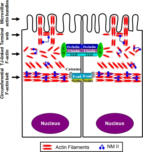
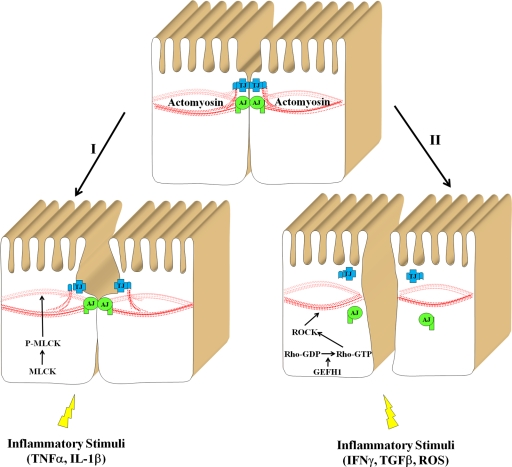
Increased epithelial permeability is a common and important consequence of mucosal inflammation that results in perturbed body homeostasis and enhanced exposure to external pathogens. The integrity and barrier properties of epithelial layers are regulated by specialized adhesive plasma membrane structures known as intercellular junctions. It is generally believed that inflammatory stimuli increase transepithelial permeability by inducing junctional disassembly. This review highlights molecular events that lead to disruption of epithelial junctions during inflammation. We specifically focus on key mechanisms of junctional regulation that are dependent on reorganization of the perijunctional F-actin cytoskeleton. We discuss critical roles of myosin-II-dependent contractility and actin filament turnover in remodeling of the F-actin cytoskeleton that drive disruption of epithelial barriers under different inflammatory conditions. Finally, we highlight signaling pathways induced by inflammatory mediators that regulate reorganization of actin filaments and junctional disassembly in mucosal epithelia.


Figures

Figure 1
Figure 1
Organization of the apical actin cytoskeleton in polarized epithelia. The apical actin cytoskeleton in polarized epithelial cells consists of several interconnecting structures that include apical microvillar actin bundles, terminal web, actomyosin meshwork associated with tight junctions, and the circumferential actomyosin belt connected to adherens junctions. Reproduced and modified with permission from Ivanov.
Figure 2
Figure 2
Mucosal inflammation in vivo induces orchestrated disruption of the perijunctional F-actin belt and TJ disassembly. Tissue sections of colonic mucosa obtained from a normal human subject and a patient with active Crohn’s disease were double-fluorescently labeled for occludin (green) and F-actin (red). Confocal microscopy images show significant colocalization (yellow) of occludin with the apical circumferential F-actin belt in normal intestinal mucosa (arrows). By contrast, occludin is relocated from TJs into the cytoplasm and F-actin belt is disrupted (arrowhead) in the intestinal epithelium of Crohn’s disease patient. Bar, 20 μm.
Figure 3
Figure 3
Signaling pathways that mediate actomyosin-dependent disruption of the epithelial barrier in inflammation. Two signaling pathways that are activated by different inflammatory stimuli and result in disruption of the epithelial barrier are presented. The first pathway involves activation of MLCK resulting in a modest contraction of perijunctional actomyosin belt and reversible increase in paracellular permeability without gross alterations in AJC structure. The second pathway involves activation of the GEF-H1-Rho-ROCK pathway that leads to profound actomyosin contraction and AJC disassembly.
Figure 4
Figure 4
Regulation of actin filament turnover. The Figure presents a simplified model of actin filament turnover and shows two mechanisms regulating the velocity of this process. The increased F-actin turnover can be mediated by actin-depolymerizing factor (ADF)/cofilin-driven F-actin depolymerization, whereas decreased turnover can be achieved by eplin and/or actinin-dependent filament cross-linking.

References

    1. Tsukita S, Furuse M, Itoh M. Multifunctional strands in tight junctions. Nat Rev Mol Cell Biol. 2001;2:285–293. - PubMed
    1. Hartsock A, Nelson WJ. Adherens and tight junctions: structure, function and connections to the actin cytoskeleton. Biochim Biophys Acta. 2008;1778:660–669. - PMC - PubMed
    1. Pokutta S, Weis WI. Structure and mechanism of cadherins and catenins in cell-cell contacts. Annu Rev Cell Dev Biol. 2007;23:237–261. - PubMed
    1. Bretscher A. Microfilament structure and function in the cortical cytoskeleton. Annu Rev Cell Biol. 1991;7:337–374. - PubMed
    1. Mooseker MS. Organization, chemistry, and assembly of the cytoskeletal apparatus of the intestinal brush border. Annu Rev Cell Biol. 1985;1:209–241. - PubMed

Publication types