. 2011 May;44(5):631-8.
doi: 10.1165/rcmb.2010-0106OC. Epub 2010 Jun 25.

Airway epithelial NF-κB activation promotes allergic sensitization to an innocuous inhaled antigen

Affiliations

Airway epithelial NF-κB activation promotes allergic sensitization to an innocuous inhaled antigen

Jennifer L Ather et al. Am J Respir Cell Mol Biol. 2011 May.

Abstract

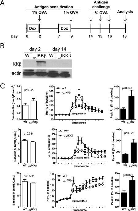
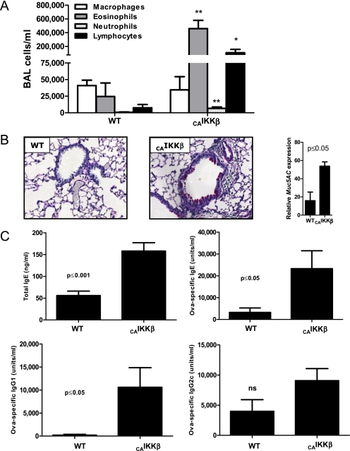
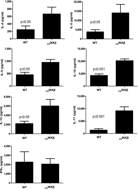
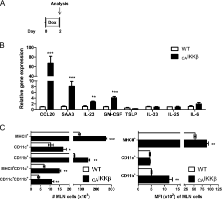
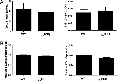
Activation of NF-κB in airway epithelium is observed in allergic asthma and is induced by inhalation of numerous infectious and reactive substances. Many of the substances that activate NF-κB in the airway epithelium are also capable of acting as adjuvants to elicit antigen-specific sensitization to concomitantly inhaled protein, thereby circumventing the inherent bias of the lung to promote tolerance to innocuous antigens. We have used a transgenic mouse inducibly expressing a constitutively active mutant of the inhibitor of nuclear factor κB (IκB) kinase β ((CA)IKKβ) that activates NF-κB only in nonciliated airway epithelial cells to test whether activation of this intracellular signaling pathway in this specific cell type is sufficient to establish a pulmonary environment permissive to the development of allergic sensitization to inhaled protein. When airway epithelial (CA)IKKβ was transiently expressed in antigen-naive mice only during initial inhalation of ovalbumin, the mice became allergically sensitized to the antigen. As a consequence, subsequent inhalation of ovalbumin alone led to an allergic asthma-like response that included airway hyperresponsiveness to methacholine, eosinophilia, mucus expression, elevated serum levels of antigen-specific IgE and IgG1, and splenic CD4(+) T cells that secreted T helper type 2 and type 17 cytokines in response to in vitro antigen restimulation. Furthermore, CD11c(+) cells in the mediastinal lymph nodes (MLN) of (CA)IKKβ-expressing mice displayed significantly elevated levels of activation markers. These data implicate airway epithelial NF-κB activation as a critical modulator of the adaptive immune response to inhaled antigens via the secretion of soluble mediators that affect the capacity of CD11c(+) cells to undergo maturation and promote antigen-specific allergic responses.


Figures

Figure 1.
Figure 1.
Airway epithelial NF-κB activation promoted antigen sensitization and airway hyperresponsiveness to methacholine. (A) A timeline of exposure regimens to promote antigen sensitization for constitutively active inhibitor of NF-κB (IκB) kinase β (CAIKKβ) transgenic and wild-type (WT) littermate mice. All 1% ovalbumin (OVA) inhalations were 30 minutes long. (B) Western blot of lung homogenates for expression of the CAIKKβ transgene at Days 2 and 14. Note that endogenous IKKβ is below the detection limit of these Western blot procedures. (C) Respiratory mechanics assessment of methacholine responsiveness using the forced oscillation technique in mice on Day 18 at baseline before administration of methacholine (left), during the methacholine challenge regimen (middle), and at the peak response to 25 mg/ml methacholine (right) (n = 5 mice/group).
Figure 2.
Figure 2.
Airway epithelial activation at the time of first encounter with antigen induces an influx of inflammatory cells subsequent to inhaled allergen challenge. CAIKKβ and WT littermate mice were exposed as depticed in Figure 1A. (A) On Day 18, differential cell counts were assessed from the bronchoalveolar lavage (BAL). Periodic acid–Schiff reactivity was assessed from histologic lung specimens visualized at 200× (B), and expression of the mucus gene Muc5AC was measured by quantitative RT-PCR and displayed relative to the levels in naive mice. (C) Serum Ig levels were measured by ELISA. Standard curves for OVA-specific IgE and IgG1 were generated from alum/OVA-sensitized BALB/cJ mouse serum, whereas nitrogen dioxide/OVA-sensitized (19) C57BL/6J serum was used to generate standards for OVA-specific IgG2c. For comparisons, values for the most concentrated standards were set to 10,000 U/ml (n = 6 mice/group). Data are representative of studies performed twice. *P ≤0.05; **P ≤0.01.
Figure 3.
Figure 3.
CAIKKβ transgene expression promotes an antigen-specific T helper (Th) 2– and Th17-skewed CD4+ T-cell phenotype. Splenic CD4+ T cells were incubated with antigen-presenting cells (APCs) and OVA for 96 hours, after which cytokines in cell culture supernatants were measured by Bio-Plex (n = 6 mice/group). Data are representative of studies performed twice.
Figure 4.
Figure 4.
Airway epithelial NF-κB expression modulates pulmonary CD11c+ APCs. (A) CAIKKβ and WT littermates fed doxycycline (Dox) chow for 48 hours and harvested immediately were (B) assessed by quantitative RT-PCR from whole lung (n = 4 mice/group). (C) Mediastinal lymph node cells were stained and analyzed by flow cytometry for APC markers. Total cell numbers in each subset were calculated (left) and median fluorescence intensity (MFI) was calculated for each of the cell markers used (right) (n = 3 mice/group). *P ≤ 0.05; **P ≤ 0.01; ***P ≤ 0.001.
Figure 5.
Figure 5.
Airway epithelial NF-κB expression does not affect epithelial barrier integrity. CAIKKβ and WT littermates fed Dox chow for 48 hours and harvested immediately. (A) BAL fluid was collected and total protein by Bradford assay (left) and lactate dehydrogenase activity (right) was recorded. (B) Whole-lung homogenates were assessed by quantitative RT-PCR for epithelial adherens junction markers zona occludens (ZO)–1 (left) and E-cadherin (right) (n = 4 mice per group). Data are representative of studies performed twice.

References

    1. Braman SS. The global burden of asthma. Chest 2006;130(1 Suppl):4S–12S. - PubMed
    1. Willart MA, Lambrecht BN. The danger within: endogenous danger signals, atopy and asthma. Clin Exp Allergy 2009;39:12–19. - PubMed
    1. Fanta CH. Asthma. N Engl J Med 2009;360:1002–1014. - PubMed
    1. Shore SA. Airway smooth muscle in asthma—not just more of the same. N Engl J Med 2004;351:531–532. - PubMed
    1. Borger P, Tamm M, Black JL, Roth M. Asthma: is it due to an abnormal airway smooth muscle cell? Am J Respir Crit Care Med 2006;174:367–372. - PubMed

Publication types

MeSH terms