. 2010 Aug 27;285(35):26852-26860.
doi: 10.1074/jbc.M110.147041. Epub 2010 Jun 25.

Toxoplasma gondii activates hypoxia-inducible factor (HIF) by stabilizing the HIF-1alpha subunit via type I activin-like receptor kinase receptor signaling

Affiliations

Toxoplasma gondii activates hypoxia-inducible factor (HIF) by stabilizing the HIF-1alpha subunit via type I activin-like receptor kinase receptor signaling

Mandi Wiley et al. J Biol Chem. .

Abstract

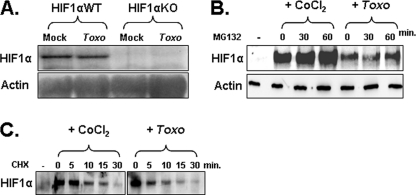
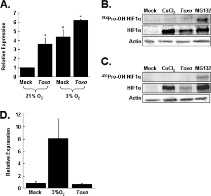
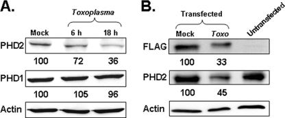
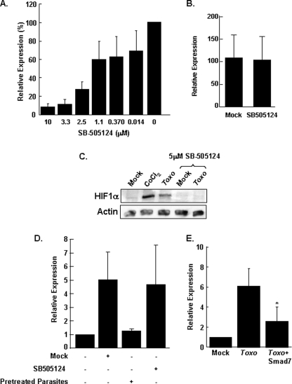
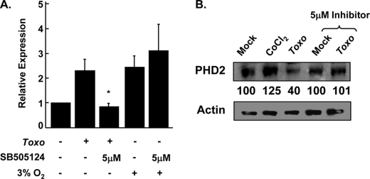
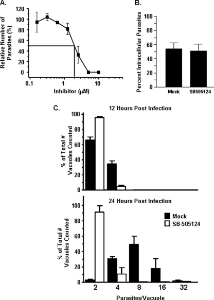
Toxoplasma gondii is an intracellular protozoan parasite that can cause devastating disease in fetuses and immune-compromised individuals. We previously reported that the alpha subunit of the host cell transcription factor, hypoxia-inducible factor-1 (HIF-1), is up-regulated by infection and necessary for Toxoplasma growth. Under basal conditions, HIF-1alpha is constitutively expressed but rapidly targeted for proteasomal degradation after two proline residues are hydroxylated by a family of prolyl hydroxylases (PHDs). The PHDs are alpha-ketoglutarate-dependent dioxygenases that have low K(m) values for oxygen, making them important cellular oxygen sensors. Thus, when oxygen levels decrease, HIF-1alpha is not hydroxylated, and HIF-1 is activated. How Toxoplasma activates HIF-1 under normoxic conditions remains unknown. Here, we report that Toxoplasma infection increases HIF-1alpha stability by preventing HIF-1alpha prolyl hydroxylation. Infection significantly decreases PHD2 abundance, which is the key prolyl hydroxylase for regulating HIF-1alpha. The effects of Toxoplasma on HIF-1alpha abundance and prolyl hydroxylase activity require activin-like receptor kinase signaling. Finally, parasite growth is severely diminished when signaling from this family of receptors is inhibited. Together, these data indicate that PHD2 is a key host cell factor for T. gondii growth and represent a novel mechanism by which a microbial pathogen subverts host cell signaling and transcription to establish its replicative niche.


Figures

FIGURE 1.
FIGURE 1.
Toxoplasma infection increases HIF-1α stability. A, total RNA from mock or Toxoplasma-infected (Toxo) cells was Northern blotted to detect HIF-1α and β-actin as a loading control. B, HFFs were mock infected, CoCl2-treated, or Toxoplasma-infected for 18 h and then treated with MG132 for the indicated times. Lysates were prepared and Western blotted to detect HIF-1α and actin as a loading control. Densitometry indicated no significant increase in HIF-1α levels in the Toxoplasma-infected cells. C, HFFs were mock infected, CoCl2-treated, or Toxoplasma-infected for 18 h and then treated with cycloheximide (CHX) for the indicated times. Lysates were prepared and Western blotted to detect anti-HIF-1α. Shown is a representative blot from three independent experiments.
FIGURE 2.
FIGURE 2.
Toxoplasma decreases prolyl hydroxylase activity. A, Renilla-ODD transfected cells were mock or parasite-infected and grown at 21 or 3% O2. Lysates were collected 24 h later, and luciferase activity was measured. Shown are the averages and standard deviations of six independent experiments. *, p < 0.05 Student's t test. B, HFFs were mock infected, CoCl2-treated, Toxoplasma-infected (Toxo) for 18 h, or MG132-treated for 2 h. C, the lysates were prepared and Western blotted with antibodies against either total HIF-1α, Pro402-OH HIF-1α, or Pro564-OH HIF-1α or actin as a loading control. Shown are representative blots from three independent experiments. D, cells cotransfected with pGBD-HIF-1α737–826 and the p5xGRE-luc reporter were either mock infected, Toxoplasma-infected, or shifted to 3% oxygen for 24 h. The lysates were collected, and luciferase activity was measured. Shown are the averages and standard deviations of three independent experiments. *, p < 0.05 Student's t test.
FIGURE 3.
FIGURE 3.
Toxoplasma decreases PHD2 protein levels. A, HFFs were mock or Toxoplasma-infected (Toxo) for 6 or 18 h. Lysates were collected and Western blotted with antibodies against PHD1, PHD2, or actin as a loading control. Shown are representative blots from three independent experiments. B, mock or FLAG-tagged PHD2-transfected HeLa cells were mock or Toxoplasma-infected for 18 h. Lysates were collected and Western blotted with antibodies to detect the FLAG epitope, PHD2, and actin as a loading control. Shown are representative blots from three independent experiments. The number listed under each band in A and B represents the amount of protein remaining relative to the mock infected samples.
FIGURE 4.
FIGURE 4.
Type I TGFβ receptor signaling is required for Toxoplasma stabilization of HIF-1α. A, pHRE-luc-transfected cells were infected in the presence of increasing concentrations of SB505124. After 24 h, the cells were lysed, and luciferase activity was measured. Shown are the means and standard deviations of three independent assays. B, pHRE-luc-transfected cells were exposed to 21 or 3% O2 in the absence or presence of SB505124. After 24 h, the cells were lysed, and luciferase activity was measured. Shown are the means and standard deviations of three independent assays. C, vehicle-treated or 5 μm SB505124-treated cells were infected with parasites or treated with CoCl2. After 18 h, the cells were lysed, and the lysates were Western blotted to detect HIF-1α or β-actin protein levels. D, mock or SB505124-treated (5 μm) pHRE-luc transfected host cells were infected with parasites, and luciferase activity was measured 18 h later. Pretreated parasites were prepared by incubating tachyzoites with 5 μm SB505124 or for 1 h and then washing the parasites with drug-free medium before adding them to pHRE-luc transfected host cells. E, pHRE-luc-transfected cells were cotransfected with either SMAD7 or empty vector as a control. The cells were mock or parasite-infected, and the luciferase activity was measured 18 h later. Shown are the averages and standard deviations from five independent experiments. *, p < 0.05 Student's t test. Toxo, Toxoplasma-infected.
FIGURE 5.
FIGURE 5.
Inhibition of ALK4,5,7 signaling blocks Toxoplasma-induced decrease in PHD2. A, vehicle- or drug-treated pTK-ODD-Rel transfected host cells were treated as indicated. Lysates were prepared 24 h later, and luciferase activity was measured. Shown are the averages and standard deviations from three independent experiments. B, vehicle-treated or 5 μm SB505124-treated cells were infected with parasites or treated with CoCl2. After 18 h, the cells were lysed, and lysates were Western blotted to detect PHD2 and β-actin protein levels. The number listed under each band represents the amount of protein remaining relative to the mock infected samples. Toxo, Toxoplasma-infected.
FIGURE 6.
FIGURE 6.
ALK4,5,7 signaling is required for Toxoplasma replication. A, parasite growth was measured 72 h after infected cells with β-galactosidase-expressing parasites in the presence of increasing levels of Type I Inhibitor. B, parasite invasion was measured by pretreating host cells with 5 μm SB505124 for 60 min. GFP-expressing parasites were then added, and the cells were fixed but not permeabilized 60 min later. Invasion was determined by differential SAG1 staining. C, parasite replication was measured by infecting mock or 5 μm SB505124-treated cells and then fixing the cells 12 or 24 h later. The parasites were detected by staining with anti-SAG1 antibody, and the numbers of parasites in individual vacuoles were counted. Shown are the averages and standard deviations from three independent experiments.

References

    1. Kim K., Weiss L. M. (2004) Int. J. Parasitol. 34, 423–432 - PMC - PubMed
    1. Montoya J. G., Liesenfeld O. (2004) Lancet 363, 1965–1976 - PubMed
    1. Blader I. J., Saeij J. P. (2009) Acta Pathol. Microbiol. Immunol. Scand. 117, 458–476 - PMC - PubMed
    1. Coppens I., Joiner K. A. (2001) Expert. Rev. Mol. Med. 2001, 1–20 - PubMed
    1. Zeiner G. M., Norman K. L., Thomson J. M., Hammond S. M., Boothroyd J. C. (2010) PLoS ONE 5, e8742. - PMC - PubMed

Publication types

MeSH terms

Substances