. 2010 Nov;38(20):7079-88.
doi: 10.1093/nar/gkq571. Epub 2010 Jun 25.

All-codon scanning identifies p53 cancer rescue mutations

Affiliations

All-codon scanning identifies p53 cancer rescue mutations

Roberta Baronio et al. Nucleic Acids Res. 2010 Nov.

Abstract

In vitro scanning mutagenesis strategies are valuable tools to identify critical residues in proteins and to generate proteins with modified properties. We describe the fast and simple All-Codon Scanning (ACS) strategy that creates a defined gene library wherein each individual codon within a specific target region is changed into all possible codons with only a single codon change per mutagenesis product. ACS is based on a multiplexed overlapping mutagenesis primer design that saturates only the targeted gene region with single codon changes. We have used ACS to produce single amino-acid changes in small and large regions of the human tumor suppressor protein p53 to identify single amino-acid substitutions that can restore activity to inactive p53 found in human cancers. Single-tube reactions were used to saturate defined 30-nt regions with all possible codon changes. The same technique was used in 20 parallel reactions to scan the 600-bp fragment encoding the entire p53 core domain. Identification of several novel p53 cancer rescue mutations demonstrated the utility of the ACS approach. ACS is a fast, simple and versatile method, which is useful for protein structure-function analyses and protein design or evolution problems.


Figures
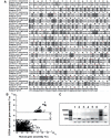
Figure 1.
Figure 1.
Self-assembly of CODA-designed human p53. (A) Alignment of the natural human p53 and CODA-optimized synthetic p53 core domain sequences. The boxed nucleotides indicate changes in the nucleotide sequence. Note that the amino acid sequence is unchanged. (B) Comparison of melting temperature (Tm) profiles of correct and incorrect hybridizations between the native human p53 DNA sequence (x axis) and the CODA-optimized p53 DNA sequence (y axis). Each point corresponds to the assembly Tm for the native (x axis) and CODA-optimized (y axis) DNA sequences for the same construction oligonucleotide or intermediate DNA fragment, as calculated using the method of Larsen et al. (34). Points above the dashed lines correspond to Tms of correct hybridizations and points below correspond to Tms of incorrect hybridizations. The correct and incorrect hybridization Tms overlap in the native sequence but are well separated in the CODA-optimized sequence, which implies that every annealing location in the CODA-optimized sequence has a globally unique thermodynamic address that is well separated from every other annealing location. (C) p53 self-assembly. Overlapping oligonucleotides were used to self-assemble fragments one to six and extended by a PCR to generate intermediate DNA fragments (lanes 1–6). The six intermediate DNA fragments were mixed together and extended by overlap extension and PCR amplification to generate the synthetic p53 core domain (lane 7).
Figure 2.
Figure 2.
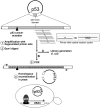
Schematic representation of the ACS strategy and the p53 cancer mutant rescue screen. A set of overlapping, degenerate oligonucleotides with roughly equal initial annealing temperatures was designed to change each single codon in a 30 bp wide region. Parallel reactions can be used to mutagenize large regions or entire genes. Parent plasmids were removed by Dpn1 digestion. At this point, a plasmid library containing completely saturated target regions of a gene of interest could be generated by transformation into E. coli. Alternatively, the mutagenized products could be amplified by PCR and introduced into target vectors by homologous recombination, or be used in many other standard applications. To select intragenic mutants that restore activity to p53 mutants found in human cancer, the p53 core domain was amplified from the mutagenized pool and introduced into a p53-tester yeast strain by homologous recombination. Yeast cells expressing re-activated p53 were identified by uracil prototrophy.
Figure 3.
Figure 3.
Representation of the experimental and theoretical frequency of codons in position 239 obtained by ACS.
Figure 4.
Figure 4.
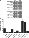
Reactivation of p53 cancer mutants by second site mutations in amino acid region 114–123. (A) Yeast cells expressing the URA3 gene from a p53-dependent promoter were transformed with centromeric plasmids (HIS3 selection marker) expressing wild-type human p53, or the mutants indicated, under control of the ADH1 promoter. Serial dilutions of cells grown to mid-log phase in medium lacking histidine were spotted onto plates either lacking histidine or uracil. Growth on plates lacking uracil is dependent on expression of the URA3 gene and is a measure of p53 activity. Mutants are named with the cancer mutation appearing first followed by the putative cancer rescue mutation. (B) p53 transcriptional activity was evaluated in transient reporter gene assays in H1299 cells, which lack endogenous p53 activity. Firefly luciferase activity, adjusted for transfection efficiency with Renilla luciferase activity, was determined 48 h after transfection. The activity of wild-type p53 was set to 100%. SD were calculated from at least three independent experiments.
Figure 5.
Figure 5.
Three-dimensional location of cancer mutants and their rescue mutations. The 3D structure of p53 (51) was visualized with Visual Molecular Dynamics (52). Positions of cancer mutations are indicated in red and positions of rescue mutations are in green.

References

    1. Jackel C, Kast P, Hilvert D. Protein design by directed evolution. Annu. Rev. Biophys. 2008;37:153–173. - PubMed
    1. Liu W, Sheppeck J.E., 2nd, Colby DA, Huang HB, Nairn AC, Chamberlin AR. The selective inhibition of phosphatases by natural toxins: the anhydride domain of tautomycin is not a primary factor in controlling PP1/PP2A selectivity. Bioorg. Med. Chem. Lett. 2003;13:1597–1600. - PubMed
    1. Yuen CM, Liu DR. Dissecting protein structure and function using directed evolution. Nat. Methods. 2007;4:995–997. - PMC - PubMed
    1. Hogrefe HH, Cline J, Youngblood GL, Allen RM. Creating randomized amino acid libraries with the quikchange multi site-directed mutagenesis kit. Biotechniques. 2002;33 1158–1160, 1162, 1164–1155. - PubMed
    1. Kegler-Ebo DM, Polack GW, DiMaio D. Use of codon cassette mutagenesis for saturation mutagenesis. Methods Mol. Biol. 1996;57:297–310. - PubMed

Publication types