. 2010 Nov 1;182(9):1153-60.
doi: 10.1164/rccm.201003-0491OC. Epub 2010 Jun 25.

Somatic chromosome abnormalities in the lungs of patients with pulmonary arterial hypertension

Affiliations

Somatic chromosome abnormalities in the lungs of patients with pulmonary arterial hypertension

Micheala A Aldred et al. Am J Respir Crit Care Med. .

Abstract

Rationale: Vascular remodeling in pulmonary arterial hypertension (PAH) involves proliferation and migration of endothelial and smooth muscle cells, leading to obliterative vascular lesions. Previous studies have indicated that the endothelial cell proliferation is quasineoplastic, with evidence of monoclonality and instability of short DNA microsatellite sequences.

Objectives: To assess whether there is larger-scale genomic instability.

Methods: We performed genome-wide microarray copy number analysis on pulmonary artery endothelial cells and smooth muscle cells isolated from the lungs of patients with PAH.

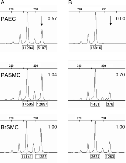
Measurements and main results: Mosaic chromosomal abnormalities were detected in PAEC cultures from five of nine PAH lungs but not in normal (n = 8) or disease control subjects (n = 5). Fluorescent in situ hybridization analysis confirmed the presence of these abnormalities in vivo in two of three cases. One patient harbored a germline mutation of BMPR2, the primary genetic cause of PAH, and somatic loss of chromosome-13, which constitutes a second hit in the same pathway by deleting Smad-8. In two female subjects with mosaic loss of the X chromosome, methylation analysis showed that the active X was deleted. One subject also showed completely skewed X-inactivation in the nondeleted cells, suggesting the pulmonary artery endothelial cell population was clonal before the acquisition of the chromosome abnormality.

Conclusions: Our data indicate a high frequency of genetically abnormal subclones within PAH lung vessels and provide the first definitive evidence of a second genetic hit in a patient with a germline BMPR2 mutation. We propose that these chromosome abnormalities may confer a growth advantage and thus contribute to the progression of PAH.


Figures

Figure 1.
Figure 1.
Chromosome abnormalities in pulmonary artery endothelial cells (PAEC) detected by single-nucleotide polymorphism (SNP) array analysis. Copy number changes can be detected on SNP arrays by a change in the locus-specific signal intensity relative to the genome-wide average and by the resulting distortion in B allele frequencies. This reduction in the chromosomal or regional copy number is presented as fractional monosomy. (A) Copy number profile of subject PAH-1 PAEC. The signal intensity for SNP probes on chromosome-13 is reduced relative to the genome-wide average, indicating a mosaic deletion. (B) Fractional monosomy in the regions of chromosome abnormality for cell lines from the 14 subjects in Table 1 (PAEC, n = 14; PASMC, n = 7; PBMC, n = 3). Statistical significance was determined using a one-sided t test, comparing the geometric mean of the candidate segment to the global mean. *Samples meeting the P value threshold of 10−7. (C) B-allele frequency distortion across the entire X chromosome in PAH-3. The value for heterozygous alleles is split into two levels, depending on the identity of the allele on the fractionally monosomic chromosome. The magnitude of this distortion is reduced in (D) PAH-4 and (E) PAH-2 in proportion to the fractional monosomy observed in these cases.
Figure 2.
Figure 2.
Fluorescent in situ hybridization analysis (FISH) of explant lung sections and cultured pulmonary artery endothelial cells (PAEC) demonstrate subject-specific chromosome monosomy in subjects PAH-3 (A, B, C) and PAH-1 (D, E, F). (A) Subject PAH-3 hematoxylin and eosin stained reference slide; (B) DAPI stained serial section with the region of intimal thickening shown in C (outlined in red); (C) FISH image of intimal region nuclei hybridized with an androgen receptor probe (Xq12, red) and CEP11 (green). Nuclei exhibiting X chromosome monosomy are indicated with yellow arrows. The proportion of cells showing a single Xq12 signal is significantly higher than those with a single chromosome-11 signal (P < 0.001; see Table 2). (D, E, F) Subject PAH-1 FISH images of cultured PAEC and lung sections hybridized with probes for RB1 (13q14.2, red), 13q34 (green), and CEP8 (aqua). PAEC metaphase (D) and interphase (E) nuclei exhibiting a single copy of chromosome-13 (yellow arrows) and two copies of chromosome-8 (white arrows). (F) Interphase FISH of a plexiform lesion within a lung section. Nuclei exhibiting chromosome 13 monosomy are indicated with yellow arrows. The proportion of cells with a single signal for both 13q14.2 and 13q34 probes is significantly higher that those with a single chromosome-8 signal (P < 0.005; see Table 2).
Figure 3.
Figure 3.
X-inactivation studies. Analysis of methylation at the androgen receptor locus in subject PAH-3. (A) Amplification of undigested DNA demonstrates a heterozygous pattern with alleles of 227bp and 233bp. Each allele is labeled with its peak height. The peak height ratio is shown, normalized to bronchial smooth muscle cells (BrSMC). Pulmonary artery smooth muscle cells (PASMC) exhibit the same peak height ratio as BrSMC, whereas in pulmonary artery endothelial cells (PAEC), the 233-bp allele (arrow) is markedly reduced in height, indicating that it is deleted in approximately 43% of cells, which is in good agreement with the microarray data (46%; see Table 1). (B) Alleles amplified from HhaI-digested DNA, where active chromatin is digested and only the inactive, methylated chromatin is PCR amplified. Peak height ratios were calculated as for panel A. PAEC show a complete absence of the 233bp allele, indicating that the 227-bp (nondeleted) allele is inactive in all cells.

Comment in

References

    1. Tuder RM, Groves B, Badesch DB, Voelkel NF. Exuberant endothelial cell growth and elements of inflammation are present in plexiform lesions of pulmonary hypertension. Am J Pathol 1994;144:275–285. - PMC - PubMed
    1. Rubin LJ. Primary pulmonary hypertension. N Engl J Med 1997;336:111–117. - PubMed
    1. Gaine SP, Rubin LJ. Primary pulmonary hypertension. Lancet 1998;352:719–725. - PubMed
    1. Moncada S, Higgs EA. Prostaglandins in the pathogenesis and prevention of vascular disease. Blood Rev 1987;1:141–145. - PubMed
    1. Moncada S. Nitric oxide. J Hypertens Suppl 1994;12:S35–S39. - PubMed

Publication types

MeSH terms

Substances