. 2010 Jun;22(6):1702-15.
doi: 10.1105/tpc.109.068627. Epub 2010 Jun 25.

VERDANDI is a direct target of the MADS domain ovule identity complex and affects embryo sac differentiation in Arabidopsis

Affiliations

VERDANDI is a direct target of the MADS domain ovule identity complex and affects embryo sac differentiation in Arabidopsis

Luis Matias-Hernandez et al. Plant Cell. 2010 Jun.

Abstract

In Arabidopsis thaliana, the three MADS box genes SEEDSTICK (STK), SHATTERPROOF1 (SHP1), and SHP2 redundantly regulate ovule development. Protein interaction studies have shown that a multimeric complex composed of the ovule identity proteins together with the SEPALLATA MADS domain proteins is necessary to determine ovule identity. Despite the extensive knowledge that has become available about these MADS domain transcription factors, little is known regarding the genes that they regulate. Here, we show that STK, SHP1, and SHP2 redundantly regulate VERDANDI (VDD), a putative transcription factor that belongs to the plant-specific B3 superfamily. The vdd mutant shows defects during the fertilization process resulting in semisterility. Analysis of the vdd mutant female gametophytes indicates that antipodal and synergid cell identity and/or differentiation are affected. Our results provide insights into the pathways regulated by the ovule identity factors and the role of the downstream target gene VDD in female gametophyte development.


Figures

Figure 1.
Figure 1.
Quantitative Real-Time PCR on Chromatin Immunoprecipitated with STK and SEP3 Antibodies. (A) Schematic representation of the position of the CArG boxes in the promoter region of the VDD gene. (B) ChIP enrichment tests by quantitative real-time PCR show STK- and SEP3-specific binding to the CArG boxes 1 and 3. The stk single mutant was used as a negative control in the STK-ChIP and wild-type leaves as negative control for the SEP3-ChIP assays. Fold enrichment was calculated over the negative controls. Error bars represent the propagated error value using three replicates (see Methods).
Figure 2.
Figure 2.
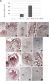
Spatial and Temporal Expression Pattern of VDD in Wild-Type, stk, shp1shp2 Double, and stk shp1 shp2 Triple Mutant Backgrounds. (A) Quantitative real-time RT-PCR performed on cDNA obtained from leaves, flower, and siliques at 3 d after pollination (DAP) and siliques at 5 DAP. The relative mRNA levels indicate that VDD is strongly expressed in the reproductive tissues before fertilization and in the early stages of seed development. Error bars represent the propagated error value using three replicates. (B) to (E) In situ hybridization experiment performed in wild-type plants. (B) VDD is expressed in the floral meristem and in developing carpels and stamens. (C) During ovule formation VDD mRNA is present in ovule primordia. (D) VDD expression is detectable during later stages of ovule formation. (E) In the mature embryo sac, VDD transcripts are found in the synergids, egg, and central cells. (F) to (H) In situ hybridization experiment performed in the stk single mutant background. (F) VDD is expressed in young flowers. (G) Within ovule primordia, the signal is reduced compared with wild-type plants. (H) Mature ovules show a decreased signal compared with wild-type plants. (I) to (K) In situ hybridization experiment performed in the shp1 shp2 double mutant background. (I) VDD is expressed in young flowers as in wild-type plants. (J) Ovule primordia in the shp1 shp2 mutant plants express VDD. (K) The hybridization signal is visible in mature ovules. (L) and (M) In situ hybridization experiment performed in the stk shp1 shp2 triple mutant background. (L) VDD is expressed in developing stamens and carpels. (M) and (N) VDD transcripts are not detected in the ovule primordia (N). No hybridization signal is visible in the carpel-like structures that develop inside the stk shp1 shp2 mutant carpel. c, carpel; cls, carpel-like structures; cc, central cell; ec, egg cell; fm, floral meristem; mo, mature ovules; op, ovule primordia; sc, synergid cells; se, sepal.
Figure 3.
Figure 3.
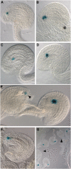
Expression of the pVDD:VDD-GUS Reporter Gene. (A) GUS expression (blue color) is detectable during ovule formation. (B) At stage 12 of flower development, GUS transcript is present in the funiculus, integuments, and nucellus. (C) Within the ovule, the VDD promoter drives the expression of the reporter gene in the gametophytic and sporophytic tissues. (D) The GUS reporter gene is transcribed in the mature female gametophyte and in the integuments. (E) Following fertilization, the selected VDD promoter was not active in the developing endosperm and embryo. c, carpel; e, embryo; es, embryo sac; f, funiculus; ii, inner integument; n, nucellus; o, ovules; oi, outer integument.
Figure 4.
Figure 4.
Seed Set in Wild-Type, vdd-1/VDD Heterozygous Plants, Complemented vdd-1 Heterozygous Plants, and pSTK:amiR-vdd Plants. (A) Schematic representation of the vdd-1 mutant allele. T-DNA is inserted in the first intron, 44 bp upstream the 3′ splicing acceptor site. (B) Wild-type silique showing full seed set. (C) Siliques of vdd-1/VDD plants containing aborted ovules (black arrows) and aborted seeds (white arrows). (D) Siliques of vdd-1/VDD plants complemented with the genomic region of the VDD gene. The complementation construct is able to rescue VDD expression in the female gametophyte. Aborted seeds (white arrowheads), but not aborted ovules, are present. (E) Siliques of wild-type plants transformed with the pSTK:amiRvdd construct. Aborted ovules only (black arrowheads) are present.
Figure 5.
Figure 5.
Fertilization Analysis in vdd-1 Heterozygous Plants. (A) Pollen tube staining with aniline blue shows that all embryo sacs in the vdd-1 heterozygous background are reached by pollen tubes. (B) Detailed image of aniline blue–stained pollen tube reaching the micropyle in the vdd-1 heterozygous carpel. (C) CLSM image of a wild-type fertilized female gametophyte. Observation performed 12 h after pollination showed one degenerating synergid in all the embryo sacs. The strong fluorescence signal indicates the degeneration of a synergid cell. (D) Detailed image of a female gametophyte in the vdd-1 heterozygous pistil. In this genetic background, not all the embryo sacs show synergid degeneration at 12 h after pollination. (E) Following pollination of wild-type pistils with pollen carrying the MINI3:GUS reporter construct, all seeds showed GUS activity in the developing endosperm. (F) Polllination of heterozygous vdd-1 pistils with MINI3:GUS pollen. GUS activity is not observed in all of the ovules. dSy, degenerated synergid; pt, pollen tube; Sy, synergid.
Figure 6.
Figure 6.
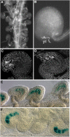
Expression Pattern of Gametophytic Cell-Specific Markers in Wild-Type and vdd-1 Heterozygous Plants. Plants homozygous for the gametophytic marker constructs were analyzed 48 HAE if not otherwise indicated. (A) and (B) Egg cell–specific marker expression. (A) Wild-type plants showed GUS expression in 98% of the female gametophytes (n = 289). (B) vdd-1 heterozygous plants showed blue staining in 97% of the megagametophytes (n = 304). (C) and (D) Central cell marker expression in wild-type plants (C) (98% of female gametophytes; n = 282) and in the vdd-1 heterozygous plants (D) (99% of megagametophytes; n = 327). (E) Antipodal cell marker expression in the vdd-1/VDD plants 72 HAE. At this time point, vdd-1/VDD heterozygous siliques showed GUS expression in the synergids cells (arrowhead) (49%; n = 244). In the remaining 51% of megagametophytes (n = 254), blue staining was visible in the antipodal cells. (F) and (G) Expression profile of the ET2634 synergid cell marker. (F) In wild-type plants, the ET2634 synergid cell marker is visible in almost all of the mature embryo sacs (96%; n = 665). (G) In the vdd-1 heterozygous plants, 32% of the mature embryo sacs (n = 740) did not express the synergid-specific cell marker (arrowheads).

Similar articles

Cited by

References

    1. Acosta-García G., Vielle-Calzada J.P. (2004). A classical arabinogalactan protein is essential for the initiation of female gametogenesis in Arabidopsis. Plant Cell 16: 2614–2628 - PMC - PubMed
    1. Battaglia R., Brambilla V., Colombo L. (2008). Morphological analysis of female gametophyte development in the bel1 stk shp1 shp2 mutant. Plant Biosyst. 142: 643–649
    1. Battaglia R., Brambilla V., Colombo L., Stuitje A.R., Kater M.M. (2006). Functional analysis of MADS-box protein complexes controlling ovule development in Arabidopsis using the ethanol-inducible alc gene-expression system. Mech. Dev. 123: 267–276 - PubMed
    1. Bemer M., Wolters-Arts M., Grossniklaus U., Angenent G.C. (2008). The MADS domain protein DIANA acts together with AGAMOUS-LIKE 80 to specify the central cell in Arabidopsis ovules. Plant Cell 208: 2088–2101 - PMC - PubMed
    1. Boisson-Dernier A., Frietsch S., Kim T.H., Dizon M.B., Schroeder J.I. (2008). The peroxin loss-of-function mutation abstinence by mutual consent disrupts male-female gametophyte recognition. Curr. Biol. 8: 63–68 - PMC - PubMed

Publication types

MeSH terms