. 2010 Oct;49(10):1852-61.
doi: 10.1093/rheumatology/keq188. Epub 2010 Jun 26.

Angiogenesis and nerve growth factor at the osteochondral junction in rheumatoid arthritis and osteoarthritis

Affiliations

Angiogenesis and nerve growth factor at the osteochondral junction in rheumatoid arthritis and osteoarthritis

David A Walsh et al. Rheumatology (Oxford). 2010 Oct.

Abstract

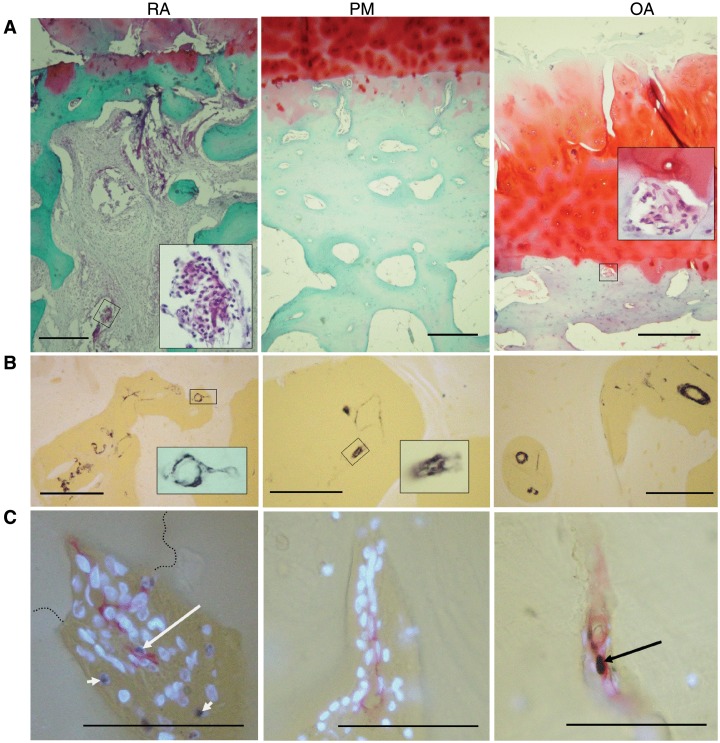
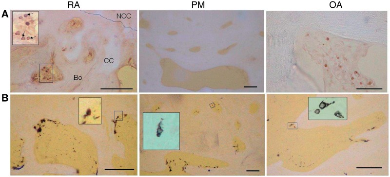
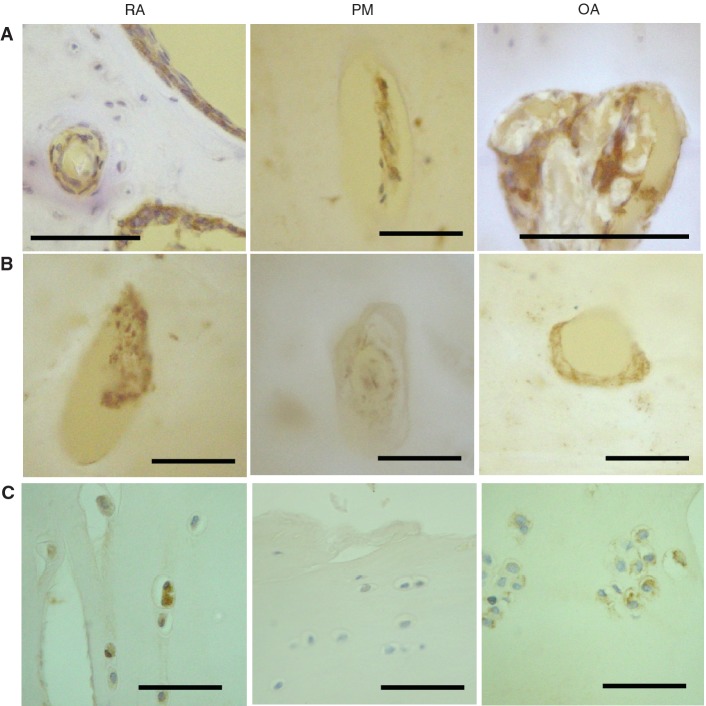
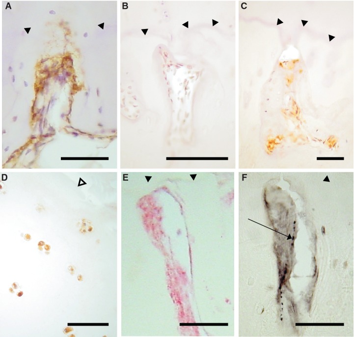
Objectives. The osteochondral junction can be a source of pain in both RA and OA. Growth of blood vessels and nerves from the subchondral bone into articular cartilage may mediate the association between joint pathology and symptoms. We have investigated associations between angiogenesis, inflammation and neurovascular growth factor expression at the osteochondral junction in human arthritis. Methods. Osteochondral junctions from medial tibial plateaux of patients undergoing arthroplasty for RA (n = 10) or OA (n = 11), or from non-arthritic post-mortem controls (n = 11) were characterized by immunohistochemistry for CD34 and smooth muscle α-actin (blood vessels), CD68 (macrophages), CD3 (lymphocytes), proliferating cell nuclear antigen, vascular endothelial, platelet-derived and nerve growth factor (NGF). Results. Osteochondral angiogenesis was demonstrated as increased endothelial cell proliferation and vascular density in non-calcified articular cartilage, both in RA and OA. Osteochondral angiogenesis was associated with subchondral bone marrow replacement by fibrovascular tissue expressing VEGF, and with increased NGF expression within vascular channels. RA was characterized by greater lymphocyte infiltration and PDGF expression than OA, whereas chondrocyte expression of VEGF was a particular feature of OA. NGF was observed in vascular channels that contained calcitonin gene-related peptide-immunoreactive sensory nerve fibres. Conclusions. Osteochondral angiogenesis in RA and OA is associated with growth factor expression by cells within subchondral spaces, vascular channels and by chondrocytes. NGF expression and sensory nerve growth may link osteochondral angiogenesis to pain in arthritis.


Figures

F<sc>ig</sc>. 1
Fig. 1
Morphology and vascularity at the osteochondral junction in RA, PM controls and OA. (A) General morphology at the osteochondral junction. RA: fibrovascular granulation tissue filling bone marrow spaces and invading the articular cartilage from below. There is proteoglycan loss and thinning of the articular cartilage and subchondral bone plate. Inset: blood vessel and inflammatory cells within the subchondral bone space. PM: normal cartilage, thick subchondral bone containing vascular channels and bone marrow spaces filled with fatty tissue. OA: fibrovascular tissue within a channel touching the tidemark (inset) as well as fibrillation, fissuring and proteoglycan depletion in cartilage at the articular surface. (B) Smooth muscle α-actin-positive cells (black) in subchondral bone spaces and associated vascular channels. Insets: positive blood vessels within a vascular channel (RA) and subchondral space (OA). (C) Vascular channels stained for CD34-positive ECs (red), PCNA-positive nuclei (black) and non-proliferating nuclei [4′-6′-diamidino-2-phenylindole (DAPI): fluorescent blue]. Proliferating ECs (long arrows) are demonstrated in a vascular channel that crosses the tidemark (broken line) in RA, and within the subchondral bone plate in OA. Proliferating non-ECs are also present (RA: short arrows). PM: vascular channel within the subchondral bone plate containing no proliferative ECs. Tissue morphology is revealed in (A) by Safranin O stain (red: proteoglycan; green: bone), with haematoxylin counterstain or (B and C) using combined transmitted and fluorescent light (blue/white: cartilage, bone and DAPI-reactive nuclei; yellow: background). Scale bars = 100  microns.
F<sc>ig</sc>. 2
Fig. 2
Inflammatory cells in subchondral bone spaces and associated vascular channels. (A) CD3-positive lymphocytes (black) within fibrovascular tissue occupying subchondral bone spaces in RA and OA, but not in the fatty marrow of a PM control. Inset: subchondral bone space showing CD3-positive lymphocytes (arrows). Bo: bone; CC: calcified cartilage; NCC: non-calcified cartilage; broken line: tidemark. (B) CD68-positive cells (black) in RA, PM controls and OA. Mononuclear CD68-positive cells resembled macrophages and were localized either at the bone surface, or deeper within the subchondral bone space. Multinucleated CD68-positive cells localized at the bone surface (insets) resembled osteoclasts. Tissue morphology is revealed by combined transmitted and fluorescent light (blue/white: cartilage, bone; yellow: background). Scale bar = 100 microns.
F<sc>ig</sc>. 3
Fig. 3
Vascular growth factor expression in articular cartilage and bone. (A) VEGF-positive cells (brown) in vascular channels (RA, PM and OA), and in bone marrow spaces (RA). (B) PDGF-positive cells (brown). Vascular channels displaying PDGF-B-positive cells within the matrix and adherent to the bone surface in RA and OA, but not PM. (C) Chondrocytes displaying VEGF immunoreactivity in deep (RA) and superficial (OA) articular cartilage, but not in PM. Tissue morphology is revealed by combined transmitted and fluorescent light (blue/white: cartilage, bone; yellow: background) (A and B) or by haematoxylin counterstain (C). Scale bar = 100 microns.
F<sc>ig</sc>. 4
Fig. 4
NGF expression in articular cartilage and bone. NGF-positive cells (brown) in vascular channels in the medial tibial plateau of patients with OA (A) and RA (C), but not in a non-arthritic control (B). (D) Chondrocytes displaying NGF immunoreactivity (brown) in superficial articular cartilage from a patient with OA (E and F). Co-localization within a vascular channel of NGF immunoreactivity (E, red) and a CGRP-immunoreactive nerve (F, black, arrow), demonstrated in serial tissue sections from a patient with OA. (A–D) DAB development with haematoxylin counterstain. (E) FastRed development. (F) Nickel-enhanced DAB development. Filled arrow heads: tidemark. Open arrow head: articular surface. Scale bar = 100 microns.

References

    1. Appel H, Kuhne M, Spiekermann S, et al. Immunohistochemical analysis of hip arthritis in ankylosing spondylitis: evaluation of the bone-cartilage interface and subchondral bone marrow. Arthritis Rheum. 2006;54:1805–13. - PubMed
    1. Bugatti S, Caporali R, Manzo A, Vitolo B, Pitzalis C, Montecucco C. Involvement of subchondral bone marrow in rheumatoid arthritis: lymphoid neogenesis and in situ relationship to subchondral bone marrow osteoclast recruitment. Arthritis Rheum. 2005;52:3448–59. - PubMed
    1. Kaneko M, Tomita T, Nakase T, et al. Expression of proteinases and inflammatory cytokines in subchondral bone regions in the destructive joint of rheumatoid arthritis. Rheumatology. 2001;40:247–55. - PubMed
    1. Lisignoli G, Piacentini A, Cristino S, et al. CCL20 chemokine induces both osteoblast proliferation and osteoclast differentiation: increased levels of CCL20 are expressed in subchondral bone tissue of rheumatoid arthritis patients. J Cell Physiol. 2007;210:798–806. - PubMed
    1. Burr DB, Schaffler MB. The involvement of subchondral mineralized tissues in osteoarthrosis: quantitative microscopic evidence. Microsc Res Tech. 1997;37:343–57. - PubMed

Publication types

Substances