. 2010 Jul;17(7):781-7.
doi: 10.1038/nsmb.1863. Epub 2010 Jun 27.

Structural and functional insights into 5'-ppp RNA pattern recognition by the innate immune receptor RIG-I

Affiliations

Structural and functional insights into 5'-ppp RNA pattern recognition by the innate immune receptor RIG-I

Yanli Wang et al. Nat Struct Mol Biol. 2010 Jul.

Abstract

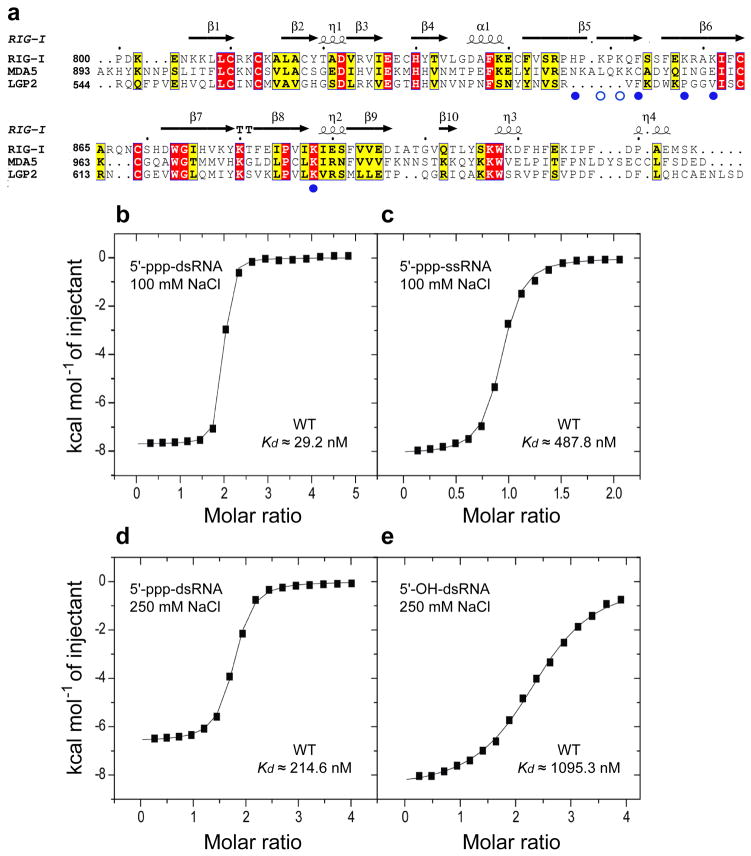
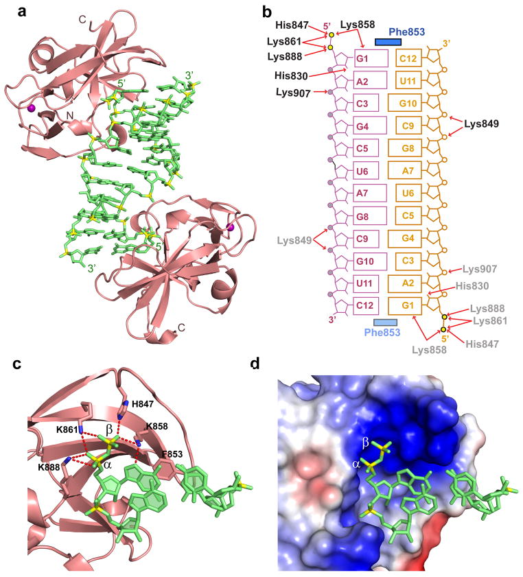
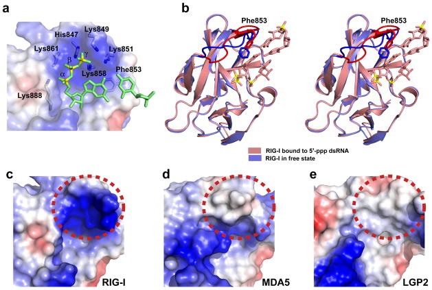
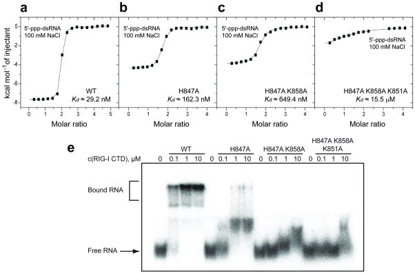
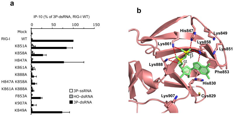
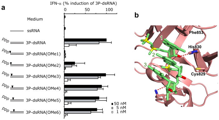
RIG-I is a cytosolic helicase that senses 5'-ppp RNA contained in negative-strand RNA viruses and triggers innate antiviral immune responses. Calorimetric binding studies established that the RIG-I C-terminal regulatory domain (CTD) binds to blunt-end double-stranded 5'-ppp RNA a factor of 17 more tightly than to its single-stranded counterpart. Here we report on the crystal structure of RIG-I CTD bound to both blunt ends of a self-complementary 5'-ppp dsRNA 12-mer, with interactions involving 5'-pp clearly visible in the complex. The structure, supported by mutation studies, defines how a lysine-rich basic cleft within the RIG-I CTD sequesters the observable 5'-pp of the bound RNA, with a stacked phenylalanine capping the terminal base pair. Key intermolecular interactions observed in the crystalline state are retained in the complex of 5'-ppp dsRNA 24-mer and full-length RIG-I under in vivo conditions, as evaluated from the impact of binding pocket RIG-I mutations and 2'-OCH(3) RNA modifications on the interferon response.


Conflict of interest statement

COMPETING INTERESTS STATEMENT

The authors declare no competing financial interests. T.T. is a co-founder and scientific advisor of Alnylam Pharmaceuticals, and also an advisor of Regulus Therapeutics.

Figures

Figure 1
Figure 1
Sequence alignment of RIG-I family of pattern recognition receptors and ITC studies of the binding of RIG-I CTD domain to RNA as a function of 5’-end phosphorylation and duplex/strand status. (a) Sequence alignment of C-terminal regulatory domains (CTDs) of RIG-I, MDA5 and LGP2. The secondary structure of RIG-I is shown over the alignments. Key amino acids contacting the α and β phosphates of the 5’-pp-dsRNA are shown by filled blue circles, while those proposed to contact the modeled γ-phosphate are shown by open blue circles. ITC binding curves for RIG-I CTD domain binding to (b) blunt-end 5’-ppp-dsRNA 12-mer and (c) single-stranded 5’-ppp-ssRNA in 100 mM NaCl and 2 mM MgCl2 buffer. ITC binding curves for RIG-I CTD domain binding to (d) blunt-end 5’-ppp-dsRNA 12-mer and (e) blunt-end 5’-OH dsRNA 12-mer in 250 mM NaCl and 2 mM MgCl2 buffer.
Figure 2
Figure 2
Details of the crystal structure of the RIG-I CTD bound to blunt-end 5’-pp-dsRNA 12-mer. (a) Crystal structure of the RIG-I CTD bound to 5’-pp-dsRNA 12-mer (in green). CTD (in salmon) are bound to both ends of the 5’-pp-dsRNA 12-mer. (b) Schematic representation highlighting intermolecular hydrogen-bonding and stacking contacts in the complex. (c) Details of the intermolecular contacts in the structure of the complex. The CTD is in a ribbon representation (salmon color) and the 5’-pp-dsRNA 12-mer is in a stick representation (green color with backbone phosphorus atoms in yellow). Intermolecular hydrogen bonds between the α and β 5’-phosphates to amino acid side chains lining the CTD recognition pocket are shown as red dotted lines. The shaded aromatic ring of F853 is stacked on both bases of the terminal base pair. (d) A view similar to panel c except for an electrostatic representation of the 5’-phosphorylated RNA-binding surface of the CTD. The RNA is in a ribbon representation and the CTD-binding pocket in an electrostatic surface representation. Blue and red patches define basic and acidic regions, respectively.
Figure 3
Figure 3
Role of 5’-phosphorylated ends in the crystal structure of the RIG-I CTD bound to blunt-end 5’-pp-dsRNA 12-mer and comparison of the electrostatics of the binding surfaces of RIG-I, MDA5 and LGP2 CTD’s for 5’-ppp-dsRNA. (a) Modeling of the γ 5’-phosphate onto the bound 5’-pp-dsRNA 12-mer in the structure of the complex. The RNA is in a ribbon representation and the CTD-binding pocket in is an electrostatic surface representation. (b) Comparison in stereo of the superposed crystal structures of the RIG-I CTD bound to blunt-end 5’-pp-dsRNA 12-mer (salmon color; only two terminal base pairs are shown in the interest of clarity) with RIG-I CTD in the free state (blue color; PDB ID: 2QFB). Conformational changes are seen for the loop spanning positions 847–856, and also in the alignment of F853, between the superposed structures and these are highlighted in darker colors. (c) The electrostatics of the binding surface of RIG-I CTD recognized by blunt-end 5’-ppp-dsRNA 12-mer. This region is shown by dashed red circle. (d) The electrostatics of the corresponding binding surface of MDA5 CTD. (e) The electrostatics of the corresponding binding surface of LGP2 CTD.
Figure 4
Figure 4
ITC and electrophoretic mobility shift studies of the binding of blunt-end 5’-ppp-dsRNA 12-mer to wild-type and mutants of RIG-I CTD. ITC binding curves for blunt-end 5’-ppp-dsRNA 12-mer binding to (a) wild-type, (b) H847A single mutant, (c) H847A/K858A double mutant and (d) H847A/K858A/K851A triple mutant of RIG-I CTD domain in 100 mM NaCl and 2 mM MgCl2 buffer. (e) Palindromic radiolabeled 5’-ppp-dsRNA 12-mer was incubated at increasing concentrations of recombinant protein under the same salt and buffer conditions as the ITC experiment. The complexes were resolved on a native polyacrylamide gel. Increasing numbers of mutation in the triphosphate-binding pocket of the protein weaken RNA binding and reduce the distinct gel shift seen for the wild-type protein to a smear. To ensure that the palindromic 5’-ppp-RNA was exclusively present as dsRNA rather than a hairpin, we pre-annealed the RNA at 5 μM strand concentration and only diluted it to 20 nM prior to the incubation with protein.
Figure 5
Figure 5
In vivo analysis of the impact of point mutants on the biological activity of RIG-I. (a) Human RIG-I (wt) or RIG-I mutants as indicated were overexpressed in human HEK293 cells and stimulated with 5 nM single-stranded (5’-ppp-ssRNA, 5’-ppp-GFP2) or double-stranded (5’-ppp-dsRNA, 5’-ppp-GFP2+AS GFP2) synthetic triphosphorylated RNA or non-modified double-stranded RNA (5’-OH-dsRNA, 5’-OH-GFP2+As GFP2). 24 hours after stimulation, IP-10 was analyzed in the supernatants of cells. Data from three independent experiments are depicted as mean values ± SEM. (b) Positions of amino acids that were mutated in the crystal structure of the 5’-pp-dsRNA 12-mer bound to RIG-I CTD.
Figure 6
Figure 6
In vivo analysis of 5’-ppp-RNA strand 2′-OCH3 substituent effects. (a) Synthetic 5’-ppp-GFP2 and the corresponding 2’-O-methyl-derivatives (5’-ppp-GFP2 OMe1 to OMe6) were hybridized with the complementary antisense strand (GFP2 AS) and transfected into human chloroquine-treated PBMCs. GFP2 AS was used as a control. IFN-α production was analyzed 20 hr after stimulation. Data from four independent donors are depicted as normalized mean values ± SEM. (b) Position of 2’-OH groups (in red) of nucleosides 1, 2 and 3 adjacent to the 5’-phosphorylated end in the crystal structure of the 5’-pp-dsRNA 12-mer bound to RIG-I CTD. Amino acids that either hydrogen bond or are in close proximity of the 2’-OH groups are also labeled in the figure.

References

    1. Takeuchi O, Akira S. Pattern recognition receptors and inflammation. Cell. 2010;140:805–820. - PubMed
    1. Yoneyama M, Fujita T. Recognition of viral nucleic acids in innate immunity. Rev Med Virol. 2010;20:4–22. - PubMed
    1. Wilkins C, Gale M., Jr Recognition of viruses by cytoplasmic sensors. Curr Opin Immunol. 2010;22:41–47. - PMC - PubMed
    1. Rehwinkel J, Reis e Sousa C. RIGorous detection: Exposing virus through RNA sensing. Science. 2010;327:284–286. - PubMed
    1. Coch C, et al. Higher activation of TLR9 in plasmacytoid dendritic cells by microbial DNA compared with self-DNA based on CpG-specific recognition of phosphodiester DNA. J Leukoc Biol. 2009;86:663–670. - PubMed

Publication types

MeSH terms

Associated data