. 2010 Jul;28(7):738-42.
doi: 10.1038/nbt.1645. Epub 2010 Jun 27.

Chemical genetics screen for enhancers of rapamycin identifies a specific inhibitor of an SCF family E3 ubiquitin ligase

Affiliations

Chemical genetics screen for enhancers of rapamycin identifies a specific inhibitor of an SCF family E3 ubiquitin ligase

Mariam Aghajan et al. Nat Biotechnol. 2010 Jul.

Abstract

The target of rapamycin (TOR) plays a central role in eukaryotic cell growth control. With prevalent hyperactivation of the mammalian TOR (mTOR) pathway in human cancers, strategies to enhance TOR pathway inhibition are needed. We used a yeast-based screen to identify small-molecule enhancers of rapamycin (SMERs) and discovered an inhibitor (SMER3) of the Skp1-Cullin-F-box (SCF)(Met30) ubiquitin ligase, a member of the SCF E3-ligase family, which regulates diverse cellular processes including transcription, cell-cycle control and immune response. We show here that SMER3 inhibits SCF(Met30) in vivo and in vitro, but not the closely related SCF(Cdc4). Furthermore, we demonstrate that SMER3 diminishes binding of the F-box subunit Met30 to the SCF core complex in vivo and show evidence for SMER3 directly binding to Met30. Our results show that there is no fundamental barrier to obtaining specific inhibitors to modulate function of individual SCF complexes.


Figures
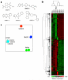
Figure 1
Figure 1. Two unsupervised data analyses classify five SMERs into three different groups based on their gene expression profiles
a, Chemical structures of SMER1 to SMER5. b, Two dimensional (2-D) hierarchical clustering reveals that the expression profile of SMER1 is similar to that of rapamycin, whereas the profiles of SMERs 2, 4 and 5 are indistinguishable from that of DMSO (solvent) control. The profile of SMER3 is distinct. Each row corresponds to a gene, and each column corresponds to an experimental sample. c, Principal component analysis is consistent with hierarchical clustering. Light blue: DMSO; blue: SMER1; cyan: SMER2; red: SMER3; sage: SMER4; chartreuse: SMER5; green: rapamycin. Replicates were obtained from independent small molecule treatments in separate experiments.
Figure 2
Figure 2. SMER3 targets SCFMet30
a, Biochemical evidence for SCFMet30 inhibition by SMER3 but not rapamycin. Yeast cells were cultured in YPDA medium to mid-log 0.8×10⁷ cells/ml, treated with indicated concentrations of SMER3 or rapamycin for 45 min, and total protein was extracted for Western blot analyses (Supplementary Information). Met4 ubiquitination in vivo can be directly assessed by immunoblotting because ubiquitinated forms of Met4 are not subjected to proteasomal degradation and can thus be detected due to a characteristic mobility shift on denaturing gels. Asterisk (*) denotes a non-specific band immuno-reactive to the anti-Met4 antibody (generous gift from Mike Tyers). b, SMER3 resistance in met4Δ cells. Yeast cells were treated with either vehicle (DMSO) or 4 μM SMER3 and growth curve analysis was performed with an automated absorbance reader measuring O.D. at 595 nm every 30 min (Supplementary Information). Cell growth was measured in liquid because SMER3 activity is undetectable on agar. c, Genetic interaction between SCFMet30 and TOR. Temperature sensitive mutants as indicated were grown at 25°C to mid-log phase in YPDA medium and serial dilutions were spotted onto plates with or without 2.5 nM rapamycin. The plates were incubated at the permissive temperatures for the mutants: 28°C for cdc34-3, cdc53-1, cdc4-3 and met30-6 because these mutants exhibited fitness defects at 30°C even without rapamycin, or 30°C (standard growth temperature) for met30-9 and skp1-25 because these alleles are not temperature sensitive until at 37°C. d, SMER3 specifically inhibits SCFMet30 E3 ligase in vitro. Components of SCFMet30 were co-expressed in insect cells and the complex was purified based on a GST-tag fused to Skp1. Met4 expressed in insect cells was bound to SCFMet30 and the ligase-substrate complex eluted with glutathione. Purified ligase-substrate complexes were combined with purified SCFCdc4 and phosphorylated Sic1 and pre-incubated with DMSO or the indicated concentrations of SMER3 for 20 min at room temperature. The ubiquitination reaction was initiated by addition of E1, E2, ubiquitin, and ATP. The reaction was allowed to proceed for 25 min, with an aliquot of the reaction collected after the first 5 min to accommodate different reaction kinetics by the two SCFs. Reaction products were analyzed by immunoblotting. The asterisks indicate a protein cross-reacting with the anti-Met4 antibody. e, The amount of un-ubiquitinated substrate (Met4 and Sic1) was quantified on a Fuji LAS-4000 imaging system and inhibition was expressed as the ratio of un-ubiquitinated substrate in DMSO/SMER3.
Figure 3
Figure 3. Molecular mechanism for the specificity of SCFMet30 inhibition by SMER3
a, Protein-protein interaction between Met30 and Skp1 is diminished by SMER3 in vivo. Yeast strains expressing endogenous 13Myc-tagged Met30 were either untreated, or treated with solvent control (DMSO) or 30μM SMER3 for 30 minutes at 30°C. 13MycMet30 was immunopurified and immuncomplexes were analyzed for Skp1 binding by Western blot analysis. b, SMER3 specifically targets SCFMet30 in vivo as determined by quantitative mass spectrometry. A yeast strain expressing endogenous HBTH-tagged Skp1 was grown in medium containing either heavy (13C/15N) or light (12C/14N) arginine and lysine to metabolically label proteins. The “heavy” culture was treated with solvent control (DMSO) and the “light” culture with 20μM SMER3 for 30 minutes at 30°C. Cells were incubated with 1% formaldehyde to cross-link and stabilize protein complexes in vivo for 10 minutes at 30°C. Cell lysates were prepared under denaturing conditions in 8M urea, mixed at equal amounts, and HBTHSkp1-bound complexes were sequentially purified on Ni2+ and streptavidin sepharose under fully denaturing conditions. Tryptic peptides of the purified complexes were analyzed by LC-MS/MS. Relative abundance of proteins was determined by measuring the peptide peak intensities. Abundance ratios for SCF components identified by multiple quantifiable peptides are shown as SILAC ratios of “light” (SMER3-treated) over “heavy” (DMSO-treated) peptide intensities. c, SMER3 specificity for SCFMet30 vs. SCFCdc4 as verified by cell cycle arrest morphology. Temperature sensitive mutants were shifted to 37°C for 4 hours. The Skp1 depletion phenotype was observed after repression of Skp1 expression in dextrose medium for 12 hours. SMER3 treatment of cells was for 6 hours. d, SMER3 directly binds to Met30-Skp1, but not Skp1 alone as determined by differential scanning fluorimetry (DSF). Met30 and Skp1 were either co-expressed or Skp1 was expressed alone in insect cells and the complex was purified based on a GST-tag fused to Met30, while Skp1 was purified based on a His-tag fused to Skp1. Protein, drug and Sypro Orange dye were added to 384-well plates and melting curve fluorescent signal was detected using the LightCycler 480 System II (Roche). Melting temperatures (Tm) were determined by the LightCycler 480 Protein Melt Analysis Tool. e, SMER3 protects endogenous Met30 from protease digestion. Yeast cells expressing Met30-RGS6H were lysed and digested with thermolysin in the presence of SMER3 vs. DMSO control, and extent of proteolysis was analyzed by immunoblotting. f, SMER3 protects recombinant Met30 from protease digestion. Met30 was PCR-subcloned into pcDNA3.1(-) (Invitrogen) and expressed using Promega TnT T7 Quick Coupled Transcription/Translation System. Thermolysin digestion was performed using translated lysate incubated with SMER3 or vehicle control, and stopped by adding EDTA pH 8.0. Samples were subjected to 4-12% NuPAGE gradient gel (Invitrogen) and Western blotted with anti-RGSH (Qiagen) and anti-GAPDH (Ambion) antibodies. The asterisks (*) indicate the Met30 fragment that is protected by SMER3 from protease digestion.

Comment in

  • Inhibitors for E3 ubiquitin ligases.
    Lydeard JR, Harper JW. Lydeard JR, et al. Nat Biotechnol. 2010 Jul;28(7):682-4. doi: 10.1038/nbt0710-682. Nat Biotechnol. 2010. PMID: 20622837 No abstract available.

References

    1. Wullschleger S, Loewith R, Hall MN. TOR signaling in growth and metabolism. Cell. 2006;124:471–484. - PubMed
    1. Bjornsti MA, Houghton PJ. The TOR pathway: a target for cancer therapy. Nat Rev Cancer. 2004;4:335–348. - PubMed
    1. Petroski MD, Deshaies RJ. Function and regulation of cullin-RING ubiquitin ligases. Nature reviews. 2005;6:9–20. - PubMed
    1. Easton JB, Houghton PJ. mTOR and cancer therapy. Oncogene. 2006;25:6436–6446. - PubMed
    1. Cloughesy TF, et al. Antitumor activity of rapamycin in a Phase I trial for patients with recurrent PTEN-deficient glioblastoma. PLoS Med. 2008;5:e8. - PMC - PubMed

Publication types

MeSH terms

Substances

Associated data