. 2010 Sep 9;29(36):5103-12.
doi: 10.1038/onc.2010.253. Epub 2010 Jun 28.

The Ras oncogene signals centrosome amplification in mammary epithelial cells through cyclin D1/Cdk4 and Nek2

Affiliations

The Ras oncogene signals centrosome amplification in mammary epithelial cells through cyclin D1/Cdk4 and Nek2

X Zeng et al. Oncogene. .

Abstract

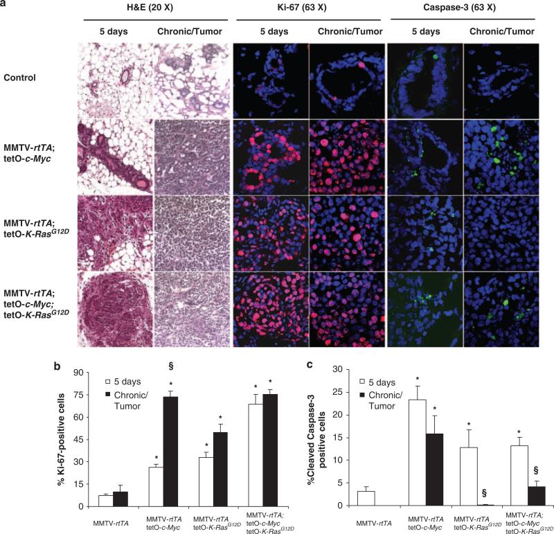
Centrosome amplification (CA) contributes to carcinogenesis by generating aneuploidy. Elevated frequencies of CA in most benign breast lesions and primary tumors suggest a causative role for CA in breast cancers. Clearly, identifying which and how altered signal transduction pathways contribute to CA is crucial to breast cancer control. Although a causative and cooperative role for c-Myc and Ras in mammary tumorigenesis is well documented, their ability to generate CA during mammary tumor initiation remains unexplored. To answer that question, K-Ras(G12D) and c-Myc were induced in mouse mammary glands. Although CA was observed in mammary tumors initiated by c-Myc or K-Ras(G12D), it was detected only in premalignant mammary lesions expressing K-Ras(G12D). CA, both in vivo and in vitro, was associated with increased expression of the centrosome-regulatory proteins, cyclin D1 and Nek2. Abolishing the expression of cyclin D1, Cdk4 or Nek2 in MCF10A human mammary epithelial cells expressing H-Ras(G12V) abrogated Ras-induced CA, whereas silencing cyclin E1 or B2 had no effect. Thus, we conclude that CA precedes mammary tumorigenesis, and interfering with centrosome-regulatory targets suppresses CA.


Figures

Figure 1
Figure 1
Inducible expression of K-RasG12D and c-Myc in mouse mammary glands results in distinct histopathology, ectopic proliferation, and apoptosis. (a) Hematoxylin–eosin (H&E) staining of mammary gland cross-sections. The first two columns show that expressions of K-RasG12D or K-RasG12D and c-Myc result in dysplasia in mice treated with doxycycline for 5 days or until tumors developed (chronic means that controls were treated chronically for 10 months but did not develop tumors). The first column was from H&E performed in paraffin-embedded 10 mm sections; the second was performed in 10 μm frozen sections. All immunostainings (columns 3–6) were performed in 7–10 μm frozen mammary cross-sections. The second pair of columns show mammary epithelial cells immunostained with antibodies against Ki-67 in red (Abcam, Cambridge, MA, USA, ab15580; the secondary antibody is conjugated with Alexa Fluor 555). The third pair of columns show mammary epithelial cells immunostained with antibodies against cleaved-caspase-3 in green (Cell Signaling, Danvers, MA, USA, 9661; the secondary antibody is conjugated with Alexa Fluor 488). The nuclei were stained with DAPI in blue. (b, c) The percentages of proliferating cells (with positive Ki-67 staining) and apoptotic cells (with positive caspase-3 staining) were calculated at 5 days or in chronically induced mice. Each group included three independent mice. Averages and s.d. were calculated as 200 epithelial cells per mouse. Significance was assessed using an unequal variance t-test, calculated in Excel (Microsoft, Redmond, WA, USA) (significance: *P<0.05 as compared with MMTV-rtTA controls, §P<0.05 when comparing the same transgenic group 5 days with long-term treatment).
Figure 2
Figure 2
Expression of K-RasG12D results in centrosome amplification in premalignant mammary lesions, whereas c-Myc-induced centrosome amplification is only detected in tumors. (a) Coimmunostaining with antibodies against γ-tubulin (Abcam, ab11317; the secondary antibody is conjugated with Alexa Fluor 555) and pericentrin (BD Biosciences, San Jose, CA, USA, 611814; the secondary antibody is conjugated with Alexa Fluor 488). Pictures presented as merged from the green and red channels, resulting in yellow signals. Arrows indicate cells with CA. Insets of the indicated area are presented for easier visualization. Chronic means that controls were treated chronically for 10 months but did not develop tumors. (b, c) Frequencies of mammary epithelial cells with 1, 2, or ≥3 centrosomes. Each group included three independent mice. Averages and s.d. were calculated as 200 epithelial cells per mouse. Significance was assessed using an unequal variance t-test, calculated in Excel (significance: *P<0.05 as compared with MMTV-rtTA controls).
Figure 3
Figure 3
Expression of K-RasG12D and c-Myc during premalignancy results in differential expression of proteins governing the cell and centrosome cycles. Lysates were extracted from the mammary glands of 3-month-old mice treated with doxycycline for 5 days. Proteins were detected by western blots. Antibodies from Cell Signaling were cyclin D1 (#2922), p16Ink4a (#4824), phospho-Ser15 p53 (#9284), phospho-Rb (Ser780, #9307; Ser807/811, #9308), Rb (#9313) and β-actin (#4970). Antibodies from BD Biosciences included Nek2 (#610593); those from Santa Cruz Biotechnology (Santa Cruz, CA, USA) included cyclin E1 (sc-481), p27Kip1 (sc-528), p21Waf1 (sc-397) and p53 (sc-6243). Duplicates (1, 2) represent two independent mice from each group.
Figure 4
Figure 4
Oncogene-induced centrosome amplification is suppressed by siRNA-mediated silencing of Cdk4, cyclin D1 or Nek2. MCF10A cells were stably transfected with plasmids encoding empty vector (pBABE-hygro or pBABE-puro), H-RasG12V (pBABE-hygro-H-RasG12V), c-Myc (pBABE-puro-c-Myc) and H-RasG12V and c-Myc (pBABE-hygro-H-RasG12V and pBABE-puro-c-Myc). These established cell lines were then transfected with control siRNAs duplexes (Ambion, Austin, TX, USA, #4611) or against cyclins B2, D1, E1, Cdk4 and Nek2 (siRNA duplexes are presented in Supplementary Table S2). (a) Western blots of cyclin B2 (Abcam, ab82287), cyclin E (Santa Cruz Biotechnology, sc-480), Cdk4 (Abcam, ab7955), cyclin D1 (Cell Signaling, 2922) and Nek2 (BD Biosciences, 610593) protein levels in MCF10A cells stably expressing vector control, H-RasG12V or H-RasG12V and c-Myc, which were starved in 0.2% FBS for 60 h. (b) Western blots of parental MCF10A transfected with control siRNAs, or siRNAs targeting cyclin B2, cyclin E1, Cdk4, cyclin D1 or Nek2, showing knockdown efficiencies. (c, d) Frequencies of CA (double immunostaining with γ-tubulin and pericentrin) and proliferation (immunostaining with BrdU, BD Pharmingen, San Jose, CA, USA, NA61) in MCF10A cells ectopically expressing empty vector, H-RasG12V, or H-RasG12V and c-Myc in the presence of control or targeted siRNAs. The average and s.d. were calculated from triplicate experiments (*P<0.05 as compared with MCF10A cells transfected with empty vector and control siRNA; §P<0.05 as compared with MCF10A cells transfected with H-RasG12V, or H-RasG12V and c-Myc vectors together with control siRNA). For each experiment, we counted at least 200 cells per group.

References

    1. Adon AM, Zeng X, Harrison MK, Sannem S, Kiyokawa H, Kaldis P, et al. Cdk2 and Cdk4 regulate the centrosome cycle and are critical mediators of centrosome amplification in p53-null cells. Mol Cell Biol. 2010;30:694–710. - PMC - PubMed
    1. Bailly E, Pines J, Hunter T, Bornens M. Cytoplasmic accumulation of cyclin B1 in human cells: association with a detergent-resistant compartment and with the centrosome. J Cell Sci. 1992;101(Part 3):529–545. - PubMed
    1. Basto R, Brunk K, Vinadogrova T, Peel N, Franz A, Khodjakov A, et al. Centrosome amplification can initiate tumorigenesis in flies. Cell. 2008;133:1032–1042. - PMC - PubMed
    1. Bearss DJ, Lee RJ, Troyer DA, Pestell RG, Windle JJ. Differential effects of p21(WAF1/CIP1) deficiency on MMTV-ras and MMTV-myc mammary tumor properties. Cancer Res. 2002;62:2077–2084. - PubMed
    1. Berman H, Zhang J, Crawford YG, Gauthier ML, Fordyce CA, McDermott KM, et al. Genetic and epigenetic changes in mammary epithelial cells identify a subpopulation of cells involved in early carcinogenesis. Cold Spring Harb Symp Quant Biol. 2005;70:317–327. - PubMed

Publication types

MeSH terms