. 2010 Sep 16;29(37):5193-203.
doi: 10.1038/onc.2010.257. Epub 2010 Jun 28.

H1047R phosphatidylinositol 3-kinase mutant enhances HER2-mediated transformation by heregulin production and activation of HER3

Affiliations

H1047R phosphatidylinositol 3-kinase mutant enhances HER2-mediated transformation by heregulin production and activation of HER3

A Chakrabarty et al. Oncogene. .

Abstract

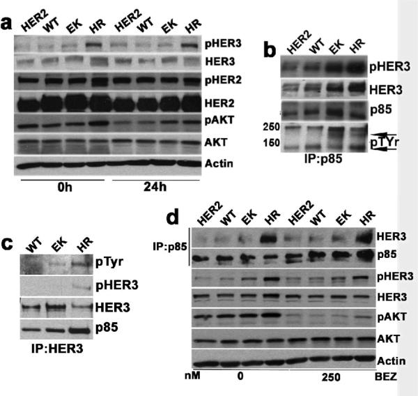
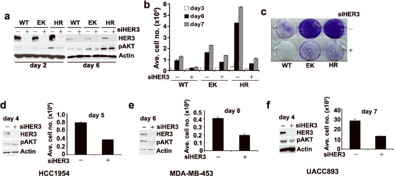
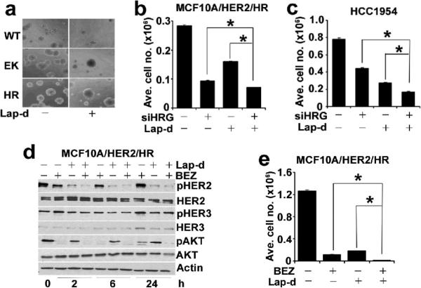
Hyperactivation of phosphatidylinositol-3 kinase (PI3K) can occur as a result of somatic mutations in PIK3CA, the gene encoding the p110α subunit of PI3K. The HER2 oncogene is amplified in 25% of all breast cancers and some of these tumors also harbor PIK3CA mutations. We examined mechanisms by which mutant PI3K can enhance transformation and confer resistance to HER2-directed therapies. We introduced the PI3K mutations E545K and H1047R in MCF10A human mammary epithelial cells that also overexpress HER2. Both mutants conferred a gain of function to MCF10A/HER2 cells. Expression of H1047R PI3K, but not E545K PI3K, markedly upregulated the HER3/HER4 ligand heregulin (HRG). HRG siRNA inhibited growth of H1047R but not E545K-expressing cells and synergized with the HER2 inhibitors trastuzumab and lapatinib. The PI3K inhibitor BEZ235 markedly inhibited HRG and pAKT levels and, in combination with lapatinib, completely inhibited growth of cells expressing H1047R PI3K. These observations suggest that PI3K mutants enhance HER2-mediated transformation by amplifying the ligand-induced signaling output of the ErbB network. This also counteracts the full effect of therapeutic inhibitors of HER2. These data also suggest that mammary tumors that contain both HER2 gene amplification and PIK3CA mutations should be treated with a combination of HER2 and PI3K inhibitors.


Figures

Figure 1
Figure 1
PI3K mutants increase transformation of MCF10A/HER2 cells. (a) Immunoblot (IB) comparing levels of HA, p110α, p85, in MCF10A/HER2 (HER2), MCF10A/HER2/WT PIK3CA (WT), MCF10A/HER2/E545K PIK3CA (EK) and MCF10A/HER2/H1047R PIK3CA (HR) cells. The HA tag was detected in cells expressing WT and mutant PI3K but not in parental MCF10A/HER2 cells. (b) IB comparing levels of total and phosphorylated AKT, S6, GSK3, total Cyclins D1 and D2 in WT, EK and HR cells. (c) 3D acinar structures of HER2, WT, EK and HR cells grown for 18 days on Matrigel ± 250f nM BEZ235 (BEZ). (d) Anchorage-independent growth of HER2, WT, EK and HR cells in soft agarose for 7 days. (e) Indirect immunofluorescence staining of cleaved caspase-3 on day 7 WT, EK and HR acini. Blue, nuclei (DAPI); green, cleaved caspase-3. (f) Transwell motility assay with WT, EK and HR cells for 24 h. (g) Invasion assay with Matrigel-coated transwell filters for 42 h.
Figure 2
Figure 2
PI3K mutants maintain pAKT and pHER3 under serum and growth factor deprivation. (a) IB comparing pHER2Y1248, pHER3Y1289 and pAKTS473 levels in HER2, WT, EK and HR cells. Cells were harvested at 0 and 24 h following serum and growth-factor deprivation. (b and c) p85-HER3 association in WT, EK and HR cells. Cell lysates were immunoprecipitated (IP) with p85 (b) and HER3 (c) antibodies, followed by IB with pTyr, HER3, pHER3Y1289 and p85 antibodies. (d) Association of pAKT, pHER3 and p85 with HER3 in cells treated with 250 nM BEZ for 6 h in growth media. IP was performed with p85 antibody. IB were performed with the antibodies indicated to the right of the panel.
Figure 3
Figure 3
RNAi of HER3 partially inhibits growth of PI3K mutant cells. (a) IB comparing HER3 and pAKT levels in cells transfected with either control or HER3 siRNA duplexes. Cells were harvested on day 2 and 6 post-transfection. (b) Determination of cell numbers on day 3, 6 and 7. Each bar represents the mean ± SE of six replicates. (c) Crystal violet staining of transfected cells on day 7. (d) HCC-1954; (e) MDA-MB-453; and (f) UACC893 cells were transfected with control or HER3 siRNA duplexes as indicated Materials and Methods. HER3 and pAKT levels were determined by IB (day 4: HCC1954; day 6: MDA-MB-453; day 4: UACC893). Differences in cell number between control and HER3 siRNA-transfected cells on day 4 for HCC1954; day 5 for MDA-MB-453 and day 7 for UACC893 were determined in a Coulter counter.
Figure 4
Figure 4
H1047R PI3K mutant cells overexpresses HER3/HER4 ligands. (a) Real-time qPCR comparing HB-EGF, EREG and HRG mRNA levels relative to housekeeping control FLJ22101 in MCF10A/HER2/WT, EK and HR cells. Each data point represents the mean ± SE of six readings. (b) IB comparing HRG levels in MCF10A/HER2/WT, EK and HR cells. (c) Comparison of pHER4 levels in MCF10A/HER2/WT, EK and HR cell lysates by IP with a HER4 antibody followed by IB with HER4, p85 and pTyr antibodies. (d) Real-time qPCR comparing HRG RNA levels in SKBR3 cells stably expressing the WT, E545K and H1047R PI3K (SKWT, SKEK and SKHR, respectively). (e) IB comparing the pHER2Y1248, pHER3Y1289, pHER3Y1222, pHER3Y1197, HER3, pAKTS473 and pERKT202 levels in SKWT, SKEK and SKHR cells treated with 1 μM Lapatinib for 0, 6 and 24 h. (f) IB comparing endogenous HRG protein levels in HER2-overexpressing cells SKBR3: WT; BT474: K111N (weakly oncogenic); HCC1954, UACC893: H1047R. MCF10A/HER2/HR cells: all harbor H1047R PI3K.
Figure 5
Figure 5
RNAi of HRG inhibits growth of H1047R but not E545K PI3K mutant cells. (a) Real-time qPCR analysis of HRG mRNA in control and HRG siRNA transfected MCF10A/HER2/HR cells. (b and c) IB comparing pHER3Y1289 (b) and pHER4Y1284 (c) levels in control and HRG siRNA transfected MCF10A/HER2/HR cells. (d) Association of p85 with HER3 and HER4 in control versus MCF10A/HER2/HR cells transfected with HRG siRNA (day 4 post-transfection). Cell lysates were precipitated with p85 antibody followed by IB with p85, HER3 and HER4 antibodies. (e) MCF10A/HER2/HR cell numbers on day 4, 5 and 6 following transfection with HRG siRNA. Each data point represents the mean ± SE of six replicates. (f) IB comparing pHER3Y1289 and HRG levels in control and HRG siRNA transfected MCF10A/HER2/EK and HR cells on day 4 post-transfection. (g) Determination of MCF10A/HER2/EK and HR cell numbers on day 7 after transfection with HRG siRNA. Each data point represents the mean ± SE of six replicates. NS, not significant; *, p<0.05, paired t-test. (h) MDA-MB-361 cells were transiently transfected with control or HRG siRNA. Cell numbers were measured in a Coulter counter on day 7 after transfection. Each data point represents the mean ± SE of six replicates. (i) IB comparing pAKTS473 levels in MCF10A/HER2/HR cells treated with vehicle (Ctrl), 20 μM LY294002 (LY) and 250 nM BEZ235 (BEZ) for 24 h. (j and k) Real-time qPCR comparing HRG mRNA levels in MCF10A/HER2/HR (j) and HCC1954 (k) cells treated or not (Ctrl) with LY and BEZ.
Figure 6
Figure 6
RNAi of HRG synergizes with HER2 TKI lapatinib. (a) 3D structures of MCF10A/HER2 cells expressing WT, EK or HR PI3K grown for 14 days on Matrigel in absence and presence of 1 μM lapatinib. b and c, MCF10A/HER2/HR (b) and HCC1954 (c) cells were transfected with control or HRG siRNAs and treated with 0.03 and 0.1 μM lapatinib, respectively. Cells numbers were measured on day 5. Each data point represents the mean ± SE of nine replicates. (d) pHER2Y1248, pHER3Y1289 and pAKTS473 in cells treated with 1 μM lapatinib and/or 250 nM BEZ by IB. (e) MCF10A/HER2/HR cells were treated with 1 μM lapatinib and/or 250nM BEZ for 6 days and cell numbers were counted. Each bar represents the mean ± SD of six replicates. *, p<0.05, paired t-test.

References

    1. Bachman KE, Argani P, Samuels Y, Silliman N, Ptak J, Szabo S, et al. The PIK3CA gene is mutated in high frequency in human breast cancers. Cancer Biol. Therapy. 2004;3:772–775. - PubMed
    1. Bader AG, Kang S, Vogt PK. Cancer-specific mutations in PIK3CA are oncogenic in vivo. Proc. Natl. Acad. Sci. USA. 2006;103:1475–1479. - PMC - PubMed
    1. Bellacosa A, Feo DD, Godwin AK, Bell DW, Cheng JQ, Altomare DA, et al. Molecular alterations of the AKT2 oncogene in ovarian and breast carcinomas. Int. J. Cancer. 1995;64:280–285. - PubMed
    1. Berns K, Horlings HM, Hennnessy BT, Mardiredjo M, Hiijmans EM, Beelen K, et al. A functional genetic approach identifies the PI3K pathway as a major determinant of trastuzuamb resistance in breast cancer. Cancer Cell. 2007;12:395–402. - PubMed
    1. Campbell IG, Russell SE, Choong DYH, Montgomery KG, Ciavarella ML, Hooi CSF, et al. Mutation in the PIK3CA gene in ovarian and breast cancer. Cancer Res. 2004;64:7678–7681. - PubMed

Publication types