. 2010 Jun 22;5(6):e11251.
doi: 10.1371/journal.pone.0011251.

Hypoxia-induced mitogenic factor (HIMF/FIZZ1/RELM alpha) recruits bone marrow-derived cells to the murine pulmonary vasculature

Affiliations

Hypoxia-induced mitogenic factor (HIMF/FIZZ1/RELM alpha) recruits bone marrow-derived cells to the murine pulmonary vasculature

Daniel J Angelini et al. PLoS One. .

Abstract

Background: Pulmonary hypertension (PH) is a disease of multiple etiologies with several common pathological features, including inflammation and pulmonary vascular remodeling. Recent evidence has suggested a potential role for the recruitment of bone marrow-derived (BMD) progenitor cells to this remodeling process. We recently demonstrated that hypoxia-induced mitogenic factor (HIMF/FIZZ1/RELM alpha) is chemotactic to murine bone marrow cells in vitro and involved in pulmonary vascular remodeling in vivo.

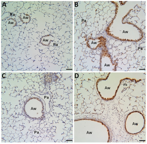
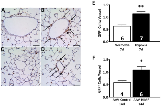
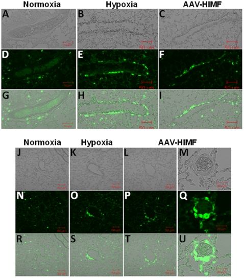
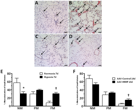
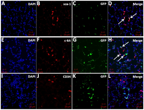
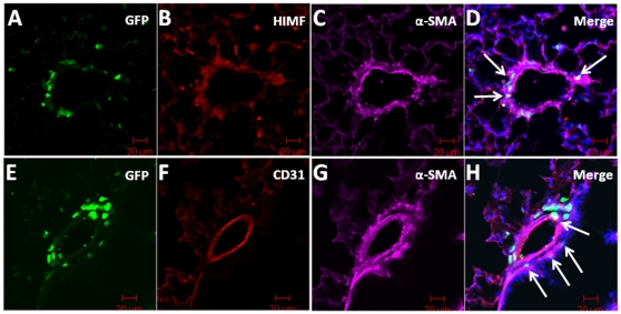
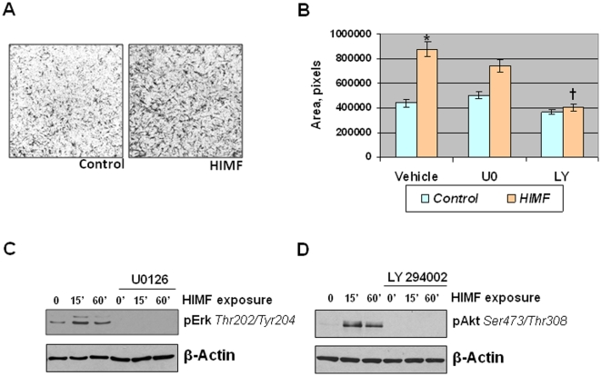
Methodology/principal findings: We used a mouse bone marrow transplant model in which lethally irradiated mice were rescued with bone marrow transplanted from green fluorescent protein (GFP)(+) transgenic mice to determine the role of HIMF in recruiting BMD cells to the lung vasculature during PH development. Exposure to chronic hypoxia and pulmonary gene transfer of HIMF were used to induce PH. Both models resulted in markedly increased numbers of BMD cells in and around the pulmonary vasculature; in several neomuscularized small (approximately 20 microm) capillary-like vessels, an entirely new medial wall was made up of these cells. We found these GFP(+) BMD cells to be positive for stem cell antigen-1 and c-kit, but negative for CD31 and CD34. Several of the GFP(+) cells that localized to the pulmonary vasculature were alpha-smooth muscle actin(+) and localized to the media layer of the vessels. This finding suggests that these cells are of mesenchymal origin and differentiate toward myofibroblast and vascular smooth muscle. Structural location in the media of small vessels suggests a functional role in the lung vasculature. To examine a potential mechanism for HIMF-dependent recruitment of mesenchymal stem cells to the pulmonary vasculature, we performed a cell migration assay using cultured human mesenchymal stem cells (HMSCs). The addition of recombinant HIMF induced migration of HMSCs in a phosphoinosotide-3-kinase-dependent manner.

Conclusions/significance: These results demonstrate HIMF-dependent recruitment of BMD mesenchymal-like cells to the remodeling pulmonary vasculature.


Conflict of interest statement

Competing Interests: The authors have declared that no competing interests exist.

Figures

Figure 1
Figure 1. HIMF expression in murine lung.
Paraffin-embedded lung sections from normoxic (7d, 20.8% O2) (A), hypoxic (7d, 10.0% O2) (B), AAV-null-treated (14d, 2.5×10¹⁰ VP) (C), and AAV-HIMF-treated (14d, 2.5×10¹⁰ VP) (D) mice were rehydrated and stained with goat anti-mouse HIMF polyclonal antibodies. Pa: pulmonary artery. Aw: airway. Scale bar: 50 µm.
Figure 2
Figure 2. Chronic hypoxia and pulmonary HIMF gene transfer increase the number of BMD cells associated with the pulmonary vasculature.
A–D: Paraffin-embedded lung sections from mice exposed to normoxia (7d, 20.8% O2) (A), hypoxia (7d, 10.0% O2) (B), AAV-null (14d, 2.5×10¹⁰ VP) (C), or AAV-HIMF (14d, 2.5×10¹⁰ VP) (D) were probed with polyclonal antibodies raised against GFP. Arrows indicate GFP+ cells within the vasculature. Scale bar: 50 µm. E, F: Quantification of GFP+ cells within the pulmonary vasculature. GFP+ cells within the pulmonary vasculature are shown as mean ± SEM of GFP+ cells/vessel. * P<0.05, ** P<0.01 vs. control.
Figure 3
Figure 3. Both chronic hypoxia and pulmonary HIMF gene transfer recruit BMD cells to the pulmonary vasculature.
(A–C, J–M) Light micrograph of fluorescence images to show blood vessel structure. Frozen sections from normoxic (20.8% O2) (D, N), hypoxic (10.0% O2) (E, O), and AAV-HIMF treated (2.5×10¹⁰ VP) (F, P, Q) lungs were stained with a rabbit anti-GFP polyclonal antibody that was visualized by an FITC-conjugated goat anti-rabbit IgG antibody (green). (G–I, R–U): Differential interference contrast images of light and fluorescence images to show structure. A–L, N–P, R–T: Scale bar: 50µm. M, Q, U: Scale bar: 20µm.
Figure 4
Figure 4. Both chronic hypoxia and pulmonary HIMF gene transfer induce pulmonary vascular remodeling.
A–D: Paraffin-embedded lung sections were double-stained with antibodies to von Willebrand factor (black) and α-smooth muscle actin (red). Arrows indicate small pulmonary vessels. Scale bar: 50 µm. E, F: Percent muscularization of small pulmonary arteries in mouse lungs. NM, non-muscularized; PM, partially muscularized; FM, fully muscularized. *Significantly decreased vs. control at P<0.05. Significantly increased vs. control at P<0.05.
Figure 5
Figure 5. GFP and cellular markers sca-1 and c-kit co-localize in AAV-HIMF-treated bone marrow transplant recipients.
Frozen lung sections from bone marrow transplant recipients treated with AAV-HIMF (2.5×10¹⁰ VP, 14d) were stained with antibodies for (B) sca-1, (F) c-kit, or (J) CD34 (red). (C, G, K) GFP signal was obtained through direct visualization (green). (A, E, I) Cell nuclei were counterstained with DAPI (blue). (D, H, L) The arrows in the merged images demonstrate co-localization of GFP with the cellular markers. Scale bar: 20 µm.
Figure 6
Figure 6. GFP+ cells were recruited to the smooth muscle layer of the pulmonary vasculature in AAV-HIMF-treated mice.
GFP was detected through direct visualization (A, E; green). HIMF and α-smooth muscle actin (α-SMA) were detected with anti-HIMF and anti-α-SMA primary antibodies and visualized with rhodamine-conjugated anti-rabbit IgG secondary antibodies (B; red) and Cy5-conjugated anti-mouse IgG secondary antibodies (C; pink), respectively. Arrows in the merged image indicate co-localization of GFP, HIMF, and α-SMA (D). Lung sections were stained with anti-CD31 antibodies and visualized with rhodamine-conjugated anti-rat IgG antibodies (F; red) and anti-α-SMA antibodies and visualized with Cy5-conjugated anti-mouse IgG antibodies (G, pink). (F) Arrows in the merged image indicate co-localization of GFP and α-SMA. Cell nuclei were stained with DAPI (D, H; blue) Scale bar: 20 µm.
Figure 7
Figure 7. HIMF-stimulated HMSC migration is PI-3K-dependent.
A: HMSCs (105 cells) were cultured in the upper chamber of a transwell plate; the lower chamber held medium containing BSA (control) or HIMF (100 nM). After 24 h, the cells were fixed and stained by Coomasie blue solution. B: HMSCs were grown as described in A, but cells were pretreated with vehicle, U0126 (10 µM), or LY294002 (10 µM). Migrated cells were quantified and results were reported as mean (±SEM) of area (pixels). *Significant increase vs. vehicle control at P<0.05. Significant decrease vs. HIMF stimulation alone at P<0.05. C, D: HMSCs were cultured to approximately 70% confluence, serum and growth factor starved overnight, and then exposed to HIMF (100 nM) or vehicle for up to 60 min in the presence or absence of ERK1/2 MAPK inhibitor U0126 (10 µM) or PI-3K inhibitor LY294002 (10 µM). Cells were lysed and proteins were resolved with SDS-PAGE and transferred to nitrocellulose membranes. The membranes were probed with rabbit anti-phospho-ERK1/2 (Thr202/Tyr204) (C) or rabbit anti-phospho-Akt (Ser473/Thr308) (D), followed by HRP-conjugated anti-rabbit IgG antibodies, and developed with ECL. To ensure equal loading and transfer, blots were stripped and reprobed with anti-β-actin.

Similar articles

Cited by

References

    1. Holcomb IN, Kabakoff RC, Chan B, Baker TW, Gurney A, et al. FIZZ1, a novel cysteine-rich secreted protein associated with pulmonary inflammation, defines a new gene family. EMBO J. 2000;19:4046–4055. - PMC - PubMed
    1. Steppan CM, Brown EJ, Wright CM, Bhat S, Banerjee RR, et al. A family of tissue-specific resistin-like molecules. Proc Natl Acad Sci U S A. 2001;98:502–506. - PMC - PubMed
    1. Teng X, Li D, Champion HC, Johns RA. FIZZ1/RELMalpha, a novel hypoxia-induced mitogenic factor in lung with vasoconstrictive and angiogenic properties. Circ Res. 2003;92:1065–1067. - PubMed
    1. Angelini DJ, Su Q, Yamaji-Kegan K, Fan C, Skinner JT, et al. Hypoxia-Induced Mitogenic Factor (HIMF) Is Involved in the Pulmonary Vascular Remodeling Associated with Chronic Hypoxia and Inflammatory Asthma. American Thoracic Society International Conference 2009
    1. Su Q, Zhou Y, Johns RA. Bruton's tyrosine kinase (BTK) is a binding partner for hypoxia induced mitogenic factor (HIMF/FIZZ1) and mediates myeloid cell chemotaxis. FASEB J. 2007;21:1376–1382. - PubMed

Publication types

Substances

LinkOut - more resources