. 2010 Apr;5(2):67-79.
doi: 10.4103/1817-1737.62469.

The calcified lung nodule: What does it mean?

Affiliations

The calcified lung nodule: What does it mean?

Ali Nawaz Khan et al. Ann Thorac Med. 2010 Apr.

Abstract

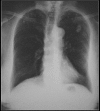
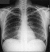
The aim of this review is to present a pictorial essay emphasizing the various patterns of calcification in pulmonary nodules (PN) to aid diagnosis and to discuss the differential diagnosis and the pathogenesis where it is known. The imaging evaluation of PN is based on clinical history, size, distribution and the gross appearance of the nodule as well as feasibility of obtaining a tissue diagnosis. Imaging is instrumental in the management of PN and one should strive not only to identify small malignant tumors with high survival rates but to spare patients with benign PN from undergoing unnecessary surgery. The review emphasizes how to achieve these goals. One of the most reliable imaging features of a benign lesion is a benign pattern of calcification and periodic follow-up with computed tomography showing no growth for 2 years. Calcification in PN is generally considered as a pointer toward a possible benign disease. However, as we show here, calcification in PN as a criterion to determine benign nature is fallacious and can be misleading. The differential considerations of a calcified lesion include calcified granuloma, hamartoma, carcinoid, osteosarcoma, chondrosarcoma and lung metastases or a primary bronchogenic carcinoma among others. We describe and illustrate different patterns of calcification as seen in PN on imaging.

Keywords: Benign pulmonary nodules; calcification; malignant pulmonary nodules.


Conflict of interest statement

Conflict of Interest: None declared.

Figures

Figure 1
Figure 1
A conventional radiograph of patient presenting with hemoptysis and weight loss shows a right hilar mass but no calcification is apparent. Two sections of axial CT scans show calcified pleural plaques (white arrows) due to previous asbestos exposure complicated by a bronchogenic neoplasm (arrow head)
Figure 2
Figure 2
It is always important to establish as to whether a PN is not from an abnormality outside the lungs. Here, a calcified nodule within the breast (arrow) mimics a PN
Figure 3
Figure 3
A chest radiograph shows a fairly well-defined PN in the right mid zone associated with a central nidus and a laminated calcification in a pulmonary hamartoma
Figure 4
Figure 4
An axial CT scan just above the hila shows a large central PN with popcorn calcification in a hamartoma
Figure 5
Figure 5
A blown-up image from a chest radiograph and axial CT scans showing low attenuation areas (arrow) within the PN due to fat virtually diagnostic of a hamartoma
Figure 6
Figure 6
Axial CT scans shows multiple small calcific PNs due to old healed histoplasmosis
Figure 7
Figure 7
Two chest radiographs 5-years apart showing a high-density solitary pulmonary nodule remaining unchanged over a 5-year period. One of the most reliable imaging features of a benign lesion is as a benign pattern of calcification and periodic follow-up with CT showing no growth for 2 years. The high density of the well-defined nodule suggest that this is calcified granuloma and no further follow-up is indicated except in patients with calcium producing tumors such as a primary osteosarcoma
Figure 8
Figure 8
A chest radiograph shows calcification at both the lung apices associated with a right lower paratracheal calcified lymph node due to healed tuberculosis
Figure 9
Figure 9
A radiograph taken for the right shoulder shows old healed granulomatous disease at the right apex of the lung associated with calcified right hilar lymph nodes
Figure 10
Figure 10
A chest radiograph and CT showing features of old healed TB. Note the loss of lung volume/fibrosis in the right upper zone and the associated pleural calcification due to a previous tuberculous empyema. Calcific granulomas are also noted in the left apical region
Figure 11
Figure 11
A chest radiograph and axial CT scan shows a dense nidus of central calcification in an adenocarcinoma of the lung
Figure 12
Figure 12
Bronchial neoplasms in contact with the thoracic wall may invade ribs and adjacent vertebrae and engulf destroyed pieces of bone and thus mimic intratumoral calcification as in this pancoast tumor
Figure 13
Figure 13
A chest radiograph of a patient with history of asbestos exposure shows multiple right-sided pleural plaques some calcified and a large pleural based PN raising the suspicion of a mesothelioma (See Figure 14)
Figure 14
Figure 14
Axial CT scans of the same patient as in Figure 13 shows a pleural-based mass with underlying pleural calcification. A CT-guided biopsy revealed a small cell lung cancer
Figure 15
Figure 15
Axial CT scan shows a central pulmonary carcinoid associated with dense amorphous calcification (arrow)
Figure 16
Figure 16
Bronchial carcinoid shown as dense nodule at the lower pole of the right hilum on the chest radiograph (arrow). On axial CT scans there is nonuniform density of the PN (white arrow), which revealed calcification on the resected specimen. Note the subsegmental atelectasis distal to the tumor (black arrow)
Figure 17
Figure 17
A Ewing's sarcoma of a rib invading the left pleura and left lung showing small high-attenuation nodules due to entrapped fragmented bone
Figure 18
Figure 18
A mediastinal/lung metastasis from a soft tissue sarcoma of the thigh showing a linear calcific density (arrow) confirmed as calcification on resected specimen
Figure 19
Figure 19
A chest radiograph showing calcified metastases from an osteogenic sarcoma. Note that the density of the tumors and the skeletal tissues is similar
Figure 20
Figure 20
PA and lateral chest radiographs showing multiple high-density lung masses in a patient with a nonmucinous adenocarcinoma of the sigmoid colon (see CT scans in Figure 21)
Figure 21
Figure 21
CT scans of the same patient as in Figure 20 shows multiple calcified metastases from a non-mucinous adenocarcinoma from sigmoid colon confirmed on CT guided needle biopsy
Figure 22
Figure 22
A chest radiograph showing a large PN at the right lung base with central high density due to calcification in a metastatic deposit from a leiomyosarcoma of the uterus
Figure 23
Figure 23
A PA chest radiograph of a patient with calcified metastases from a medullary carcinoma of the thyroid. A radiograph of the upper abdomen on the same patient showing calcified metastases to the liver
Figure 24
Figure 24
A rare calcified intralobar sequestration; the arterial supply and venous drainage is elegantly shown by the CT angiography (right) note that the arterial blood supply is arising from the celiac axis (white arrows) and venous drainage is via the left renal vein (gray arrow)
Figure 25
Figure 25
Chest radiograph and an axial CT scan shows calcification PMF in a coal miners lung
Figure 26
Figure 26
Metastatic calcification in the lungs in a patient with chronic renal failure
Figure 27
Figure 27
Miliary diffuse calcific nodules in an adult male with a previous history of Varicella pneumonia
Figure 28
Figure 28
A chest radiograph shows multiple small calcific nodules in an adult female with a past history of Varicella pneumonia
Figure 29
Figure 29
Hydatid cysts do not normally calcify within the lungs; two calcified hydatid cysts in the superior and posterior mediastinum are seen. Note the calcified hydatid cyst within the left lobe of the liver on the coronal CT reconstruction
Figure 30
Figure 30
A chest radiograph shows reticulonodular shadowing with bilateral apical lung fibrosis and high density nodules in coal workers pneumoconiosis
Figure 31
Figure 31
Two chest radiographs from the same Iron ore worker 10 years apart shows fine high-density nodules (left) progressing to PMF 10 years later
Figure 32
Figure 32
A chest radiograph shows multiple high density nodules due to a lifetime exposure to hematite and silica
Figure 33
Figure 33
Multiple calcified pleural plaques mimicking PNs on a chest radiographs elegantly depicted on axial CT scan as calcified pleural plaques from previous asbestos exposure
Figure 34
Figure 34
HRCT showing features of pulmonary alveolar microlithiasis. Note the innumerable calcific nodules bilaterally throughout both lungs giving rise to a sand-like appearance
Figure 35
Figure 35
A chest radiograph and coronal reconstruction of CT shows tubular opacities of metallic density due to acrylic cement pulmonary emboli as a complication of vertebroplasty
Figure 36
Figure 36
An axial CT of the same patient as in Figure 34 showing tubular opacities of metallic density due to acrylic cement pulmonary embolism as a complication of vertebroplasty
Figure 37
Figure 37
Section of an HRCT showing amiodarone lung. N ote the high density lung, septal thickening and features of interstitial fibrosis
Figure 38
Figure 38
A chest radiograph shows thorotrast deposition in posterior mediastinal lymph nodes seen as tiny metallic nodules. This patient had a carotid angiogram performed in the mid forties for a subarachnoid hemorrhage where thorotrast was used as a contrast agent. The spleen, which is not clearly depicted here also showed lace-like metallic densities due to thorotrast deposition

Similar articles

Cited by

References

    1. Grewal RG, Austin JH. CT demonstration of calcification in carcinoma of the lung. J Comput Assist Tomogr. 1994;18:867–71. - PubMed
    1. Caskey CI, Templeton PA, Zerhouni EA. Current evaluation of the solitary pulmonary nodule. Radiol Clin North Am. 1990;28:511–20. - PubMed
    1. Mahoney MC, Shipley RT, Corcoran HL, Dickson BA. CT demonstration of calcification in carcinoma of the lung. AJR Am J Roentgenol. 1990;154:255–8. - PubMed
    1. Siegelman SS, Khouri NF, Scott WW, Jr, Leo FP, Hamper UM, Fishman EK, et al. Pulmonary hamartoma: CT findings. Radiology. 1986;160:313–7. - PubMed
    1. Oldham HN, Jr, Young WG, Jr, Sealy WC. Hamartoma of the lung. J Thorac Cardiovasc Surg. 1967;53:735–42. - PubMed