

Co-expression of Argonaute2 Enhances Short Hairpin RNA-induced RNA Interference in Xenopus CNS Neurons In Vivo
- PMID: 20582287
- PMCID: PMC2858607
- DOI: 10.3389/neuro.17.001.2009
Co-expression of Argonaute2 Enhances Short Hairpin RNA-induced RNA Interference in Xenopus CNS Neurons In Vivo
Abstract
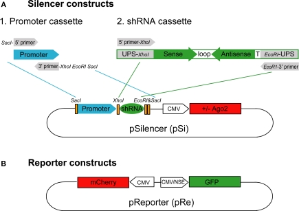
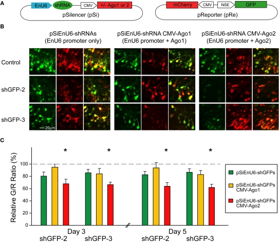
RNA interference (RNAi) is an evolutionarily conserved mechanism for sequence-specific gene silencing. Recent advances in our understanding of RNAi machinery make it possible to reduce protein expression by introducing short hairpin RNA (shRNA) into cells of many systems, however, the efficacy of RNAi-mediated protein knockdown can be quite variable, especially in intact animals, and this limits its application. We built adaptable molecular tools, pSilencer (pSi) and pReporter (pRe) constructs, to evaluate the impact of different promoters, shRNA structures and overexpression of Ago2, the key enzyme in the RNA-induced silencing complex, on the efficiency of RNAi. The magnitude of RNAi knockdown was evaluated in cultured cells and intact animals by comparing fluorescence intensity levels of GFP, the RNAi target, relative to mCherry, which was not targeted. Co-expression of human Ago2 with shRNA significantly enhanced efficiency of GFP knockdown in cell lines and in neurons of intact Xenopus tadpoles. Human H1- and U6-promotors alone or the U6-promotor with an enhancer element were equally effective at driving GFP knockdown. shRNA derived from the microRNA-30 design (shRNA(mir30)) enhanced the efficiency of GFP knockdown. Expressing pSi containing Ago2 with shRNA increased knockdown efficiency of an endogenous neuronal protein, the GluR2 subunit of the AMPA receptor, functionally accessed by recording AMPA receptor-mediated spontaneous synaptic currents in Xenopus CNS neurons. Our data suggest that co-expression of Ago2 and shRNA is a simple method to enhance RNAi in intact animals. While morpholino antisense knockdown is effective in Xenopus and Zebrafish, a principle advantage of the RNAi method is the possibility of spatial and temporal control of protein knockdown by use of cell type specific and regulatable pol II promoters to drive shRNA and Ago2. This should extend the application of RNAi to study gene function of intact brain circuits.
Keywords: AMPA receptor; Ago2; Pol III promoter; RNAi; Xenopus; knockdown; shRNA.
Figures


Similar articles
-
A new design of a lentiviral shRNA vector with inducible co-expression of ARGONAUTE 2 for enhancing gene silencing efficiency.Cell Biosci. 2015 Dec 8;5:67. doi: 10.1186/s13578-015-0058-2. eCollection 2015. Cell Biosci. 2015. PMID: 26649169 Free PMC article.
-
Construction of simple and efficient DNA vector-based short hairpin RNA expression systems for specific gene silencing in mammalian cells.Methods Mol Biol. 2007;408:223-41. doi: 10.1007/978-1-59745-547-3_13. Methods Mol Biol. 2007. PMID: 18314586
-
Characterization and comparison of chicken U6 promoters for the expression of short hairpin RNAs.Anim Biotechnol. 2007;18(3):153-62. doi: 10.1080/10495390600867515. Anim Biotechnol. 2007. PMID: 17612838
-
Post-transcriptional gene silencing by RNA interference in non-mammalian vertebrate systems: where do we stand?Mutat Res. 2011 Nov-Dec;728(3):158-71. doi: 10.1016/j.mrrev.2011.09.001. Epub 2011 Sep 13. Mutat Res. 2011. PMID: 21930237 Review.
-
Short hairpin RNA-mediated gene silencing.Methods Mol Biol. 2013;942:205-32. doi: 10.1007/978-1-62703-119-6_12. Methods Mol Biol. 2013. PMID: 23027054 Review.
Cited by
-
The REMOTE-control system: a system for reversible and tunable control of endogenous gene expression in mice.Nucleic Acids Res. 2017 Dec 1;45(21):12256-12269. doi: 10.1093/nar/gkx829. Nucleic Acids Res. 2017. PMID: 28981717 Free PMC article.
-
Regulation of photoreceptor gene expression by the retinal homeobox (Rx) gene product.Dev Biol. 2010 Mar 15;339(2):494-506. doi: 10.1016/j.ydbio.2009.12.032. Epub 2010 Jan 7. Dev Biol. 2010. PMID: 20060393 Free PMC article.
-
Xenopus tropicalis as a model organism for genetics and genomics: past, present, and future.Methods Mol Biol. 2012;917:3-15. doi: 10.1007/978-1-61779-992-1_1. Methods Mol Biol. 2012. PMID: 22956079 Free PMC article.
-
A new design of a lentiviral shRNA vector with inducible co-expression of ARGONAUTE 2 for enhancing gene silencing efficiency.Cell Biosci. 2015 Dec 8;5:67. doi: 10.1186/s13578-015-0058-2. eCollection 2015. Cell Biosci. 2015. PMID: 26649169 Free PMC article.
-
Robust RNAi enhancement via human Argonaute-2 overexpression from plasmids, viral vectors and cell lines.Nucleic Acids Res. 2013 Nov;41(21):e199. doi: 10.1093/nar/gkt836. Epub 2013 Sep 17. Nucleic Acids Res. 2013. PMID: 24049077 Free PMC article.
References
Grants and funding
LinkOut - more resources
Full Text Sources
Other Literature Sources