. 2010 Jun 22;4(6):e724.
doi: 10.1371/journal.pntd.0000724.

Isolation and characterization of new Leptospira genotypes from patients in Mayotte (Indian Ocean)

Affiliations

Isolation and characterization of new Leptospira genotypes from patients in Mayotte (Indian Ocean)

Pascale Bourhy et al. PLoS Negl Trop Dis. .

Abstract

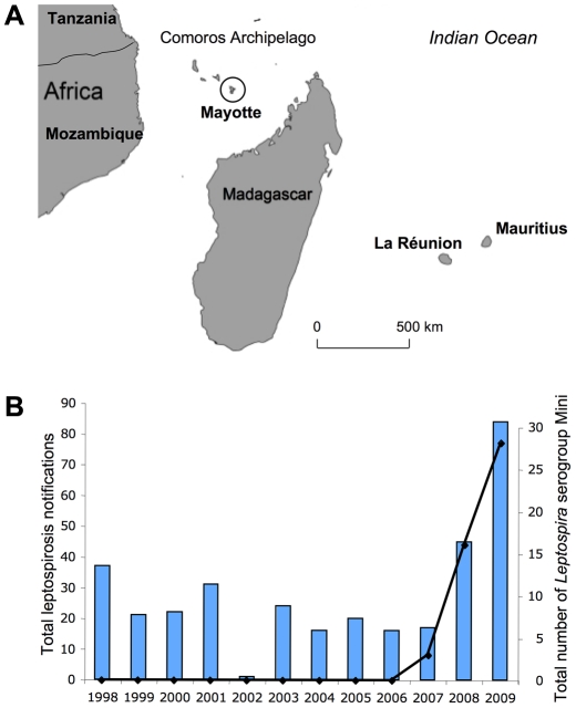
Background: Leptospirosis has been implicated as a severe and fatal form of disease in Mayotte, a French-administrated territory located in the Comoros archipelago (southwestern Indian Ocean). To date, Leptospira isolates have never been isolated in this endemic region.

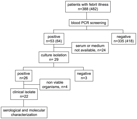
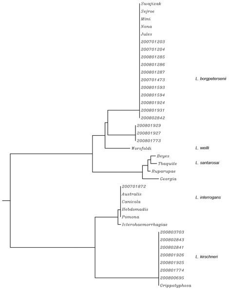
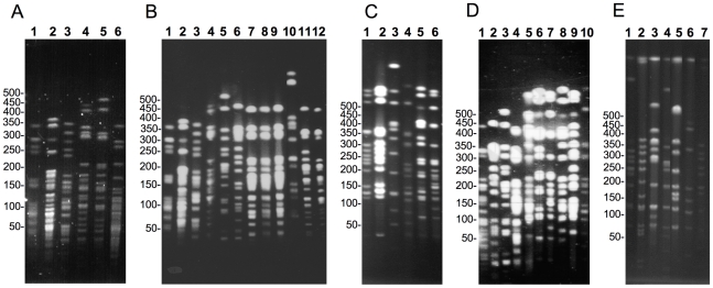
Methods and findings: Leptospires were isolated from blood samples from 22 patients with febrile illness during a 17-month period after a PCR-based screening test was positive. Strains were typed using hyper-immune antisera raised against the major Leptospira serogroups: 20 of 22 clinical isolates were assigned to serogroup Mini; the other two strains belonged to serogroups Grippotyphosa and Pyrogenes, respectively. These isolates were further characterized using partial sequencing of 16S rRNA and ligB gene, Multi Locus VNTR Analysis (MLVA), and pulsed field gel electrophoresis (PFGE). Of the 22 isolates, 14 were L. borgpetersenii strains, 7 L. kirschneri strains, and 1, belonging to serogoup Pyrogenes, was L. interrogans. Results of the genotyping methods were consistent. MLVA defined five genotypes, whereas PFGE allowed the recognition of additional subgroups within the genotypes. PFGE fingerprint patterns of clinical strains did not match any of the patterns in the reference strains belonging to the same serogroup, suggesting that the strains were novel serovars.

Conclusions: Preliminary PCR screening of blood specimen allowed a high isolation frequency of leptospires among patients with febrile illness. Typing of leptospiral isolates showed that causative agents of leptospirosis in Mayotte have unique molecular features.


Conflict of interest statement

The authors have declared that no competing interests exist.

Figures

Figure 1
Figure 1. Leptospirosis in Mayotte.
A. Location of Mayotte (circle) in the Comoros Archipelago. B. Distribution of cases of serogroup Mini compared with the total number of leptospirosis cases in Mayotte between 1998–2009.
Figure 2
Figure 2. Flow diagram showing blood sample processing.
In brackets the total number of serum samples, including serial samples.
Figure 3
Figure 3. ligB gene sequence-based phylogeny of clinical isolates from Mayotte and representative serovars belonging to serogroups Mini, Hebdomadis, and Grippotyphosa.
L. interrogans reference strains (serovars Australis, Icterohaemorragiae, Canicola, and Pomona) were also included in the dendrogram. The dendrogram was constructed by using the neighbour-joining method.
Figure 4
Figure 4. PFGE separation of NotI-digested genomic DNA from Leptospira isolates.
A. Reference strains from serogroup Mini: serovars Beye (1), Georgia (2), Ruparupae (3), Mini (4), Swajizak (5), and Tbaquite (6). B. serovar Beye (1), serovar Georgia (2), serovar Ruparupae (3), serovar Mini (4), 2008/01927 (5), 2008/01929 (6), 2008/01931 (7), 2008/02842 (8), 2008/01924 (9), serovar Kambale (10), serovar Nona (11), and serovar Jules (12). C. serovar Grippotyphosa strain Valbuzzi (1), serovar Grippotyphosa strain Moskva V (2), serovar Grippotyphosa strain Andaman (3), 2008/01774 (4), 2008/03703 (5), 2008/00695 (6). D. serovar Jules (1), 2008/01773 (2), 2008/01927 (3), 2008/01926 (4), 2008/02843 (5), 2008/03703 (6), 2008/00695 (7), 2008/01925 (8), 2008/02841 (9), 2008/01774 (10). E. 2007/01872 (1), serovar Abramis (2), serovar Biggis (3), serovar Camlo (4), serovar Guaratuba (5), serovar Manilae (6), and serovar Pyrogenes (7). The molecular weight size marker consisted of bacteriophage lambda DNA concatemers of 50 kb.
Figure 5
Figure 5. PCR analysis of the polymorphism of VNTR4, VNTR7, and VNTR10 loci of “Grippotyphosa” strains.
Lanes 1: serovar Muelleri, 2: serovar Liangguang, 3: serovar Ratnapura, 4: serovar Vanderhoedeni, 5: serovar Grippotyphosa strain DF, 6: serovar Grippotyphosa strain Moskva V., 7: strain 2008/01774.
Figure 6
Figure 6. Virulence of clinical isolate 2007/01203 in the gerbil model of leptospirosis.
Infection experiments with strain 2007/01203 were performed with four gerbils per group. Groups of gerbils were inoculated with 106, 104, 103, 102, 101, and 100 leptospires.
Figure 7
Figure 7. Histopathological sections of liver (A, D), kidney (B, E), and lung (C, F) samples from gerbils infected with clinical isolate 2007/01203.
Left panel: Hematoxylin and eosin staining (×100). Right panel: Whartin Starry staining (×1000). Hepatocyte necrosis (A, arrowhead) and Kupffer cells surrounded by polymorphonuclear and mononuclear cells were observed within the medio lobular zones of the liver (A, arrows). Periportal infiltrates with lymphocytes were also observed. Several spirochetes were distributed around the hepatocytes (D, arrow). Kidneys exhibited hemorrhaging around the tubules (B, arrowhead), necrosis of tubular epithelia, and interstitial nephritis with infiltrates of lymphocytes, and monocytes, and occasional polymorphonuclear cells (B, arrow). Spirochetes were observed within renal tubules, venular endothelia (E, arrow), and glomeruli. Infiltrates with polymorphonuclear and mononuclear cells were seen in alveolar septa (C, arrow). Spirochetes were found in the capillary network of the alveolar septa (F, arrow).

Similar articles

Cited by

References

    1. Levett PN. Leptospirosis. Clin Microbiol Rev. 2001;14:296–326. - PMC - PubMed
    1. McBride AJ, Athanazio DA, Reis MG, Ko AI. Leptospirosis. Curr Opin Infect Dis. 2005;18:376–386. - PubMed
    1. Kuriakose M, Paul R, Joseph MR, Sugathan S, Sudha TN. Leptospirosis in a midland rural area of Kerala State. Indian J Med Res. 2008;128:307–312. - PubMed
    1. Lacerda HG, Monteiro GR, Oliveira CC, Suassuna FB, Queiroz JW, et al. Leptospirosis in a subsistence farming community in Brazil. Trans R Soc Trop Med Hyg. 2008;102:1233–1238. - PMC - PubMed
    1. Ko AI, Galvao Reis M, Ribeiro Dourado CM, Johnson WDJ, Riley LW. Urban epidemic of severe leptospirosis in Brazil. Salvador Leptospirosis Study Group. Lancet. 1999;354:820–825. - PubMed

Publication types

MeSH terms