

Anatomical global spatial normalization
- PMID: 20582489
- PMCID: PMC2945458
- DOI: 10.1007/s12021-010-9074-x
Anatomical global spatial normalization
Abstract
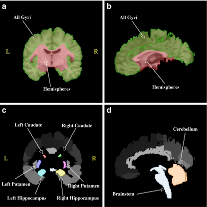
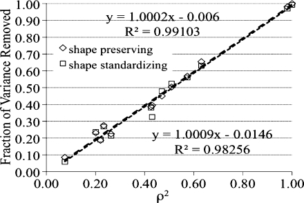
Anatomical global spatial normalization (aGSN) is presented as a method to scale high-resolution brain images to control for variability in brain size without altering the mean size of other brain structures. Two types of mean preserving scaling methods were investigated, "shape preserving" and "shape standardizing". aGSN was tested by examining 56 brain structures from an adult brain atlas of 40 individuals (LPBA40) before and after normalization, with detailed analyses of cerebral hemispheres, all gyri collectively, cerebellum, brainstem, and left and right caudate, putamen, and hippocampus. Mean sizes of brain structures as measured by volume, distance, and area were preserved and variance reduced for both types of scale factors. An interesting finding was that scale factors derived from each of the ten brain structures were also mean preserving. However, variance was best reduced using whole brain hemispheres as the reference structure, and this reduction was related to its high average correlation with other brain structures. The fractional reduction in variance of structure volumes was directly related to ρ (2), the square of the reference-to-structure correlation coefficient. The average reduction in variance in volumes by aGSN with whole brain hemispheres as the reference structure was approximately 32%. An analytical method was provided to directly convert between conventional and aGSN scale factors to support adaptation of aGSN to popular spatial normalization software packages.
Figures

Similar articles
-
Automated analysis of fundamental features of brain structures.Neuroinformatics. 2011 Dec;9(4):371-80. doi: 10.1007/s12021-011-9108-z. Neuroinformatics. 2011. PMID: 21360205 Free PMC article.
-
MR imaging volumetry of subcortical structures and cerebellar hemispheres in normal persons.AJNR Am J Neuroradiol. 2003 Apr;24(4):644-7. AJNR Am J Neuroradiol. 2003. PMID: 12695196 Free PMC article.
-
Repeatability of measured brain volume by atlas-based method using T1-weighted image.J Digit Imaging. 2012 Feb;25(1):173-8. doi: 10.1007/s10278-011-9412-z. J Digit Imaging. 2012. PMID: 21773867 Free PMC article.
-
Advances in functional imaging of the human cerebellum.Curr Opin Neurol. 2010 Aug;23(4):382-7. doi: 10.1097/WCO.0b013e32833be837. Curr Opin Neurol. 2010. PMID: 20581682 Review.
-
Computational analysis of cerebral cortex.Neuroradiology. 2010 Aug;52(8):691-8. doi: 10.1007/s00234-010-0715-4. Epub 2010 May 18. Neuroradiology. 2010. PMID: 20480153 Review.
Cited by
-
Three-Dimensional Probabilistic Maps of Mesial Temporal Lobe Structures in Children and Adolescents' Brains.Front Neuroanat. 2018 Nov 15;12:98. doi: 10.3389/fnana.2018.00098. eCollection 2018. Front Neuroanat. 2018. PMID: 30498435 Free PMC article.
-
Bayes estimate of primary threshold in clusterwise functional magnetic resonance imaging inferences.Stat Med. 2021 Nov 10;40(25):5673-5689. doi: 10.1002/sim.9147. Epub 2021 Jul 26. Stat Med. 2021. PMID: 34309050 Free PMC article.
-
Neural Mechanisms of Acceptance and Commitment Therapy for Chronic Pain: A Network-Based fMRI Approach.Front Hum Neurosci. 2021 Feb 5;15:587018. doi: 10.3389/fnhum.2021.587018. eCollection 2021. Front Hum Neurosci. 2021. PMID: 33613207 Free PMC article.
-
Scale normalization of histopathological images for batch invariant cancer diagnostic models.Annu Int Conf IEEE Eng Med Biol Soc. 2012;2012:4406-9. doi: 10.1109/EMBC.2012.6346943. Annu Int Conf IEEE Eng Med Biol Soc. 2012. PMID: 23366904 Free PMC article.
-
The brain, obesity and addiction: an EEG neuroimaging study.Sci Rep. 2016 Sep 23;6:34122. doi: 10.1038/srep34122. Sci Rep. 2016. PMID: 27658351 Free PMC article.
References
-
- Alpert NM, Bradshaw JF, Kennedy D, Correia JA. The principal axis transformation—A method for image registration. Journal of Nuclear Medicine. 1990;31:1717–1722. - PubMed
Publication types
MeSH terms
Grants and funding
LinkOut - more resources
Full Text Sources
Other Literature Sources
Medical
