. 2010 Aug;341(2):289-97.
doi: 10.1007/s00441-010-0997-1. Epub 2010 Jun 26.

Pseudopod-like basal cell processes in intestinal cholecystokinin cells

Affiliations

Pseudopod-like basal cell processes in intestinal cholecystokinin cells

Rashmi Chandra et al. Cell Tissue Res. 2010 Aug.

Abstract

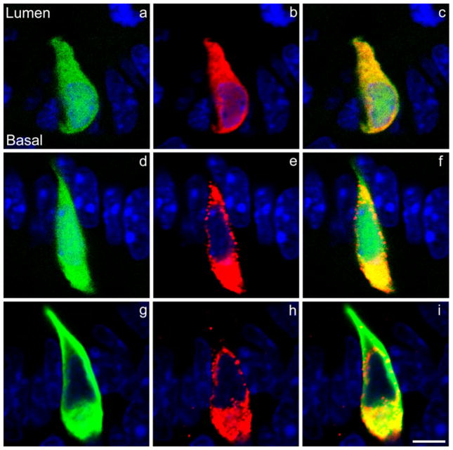
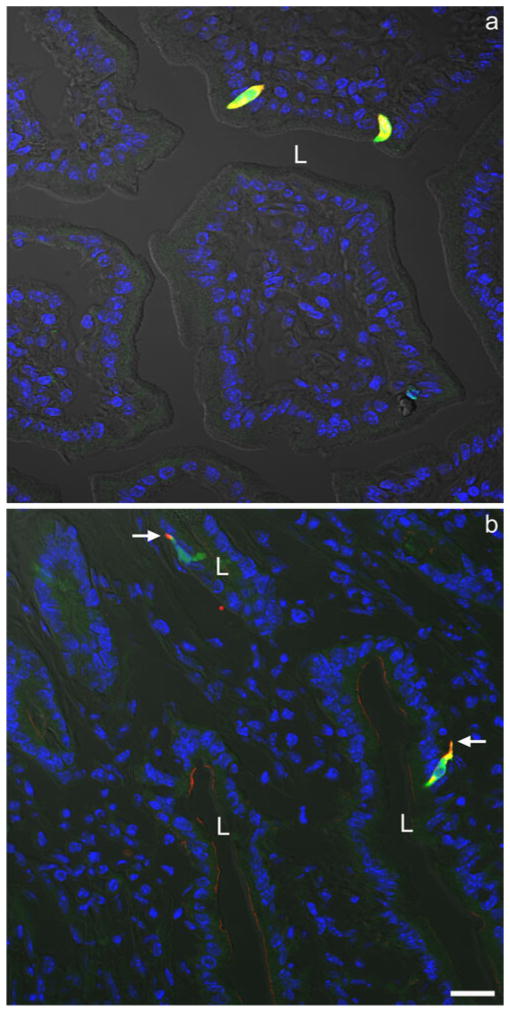
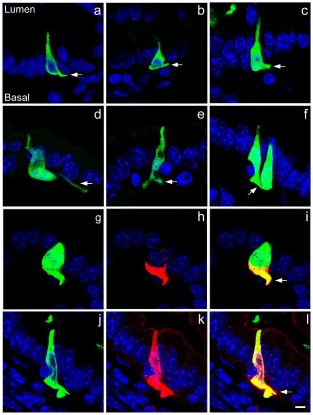
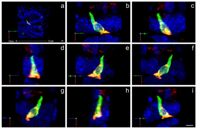
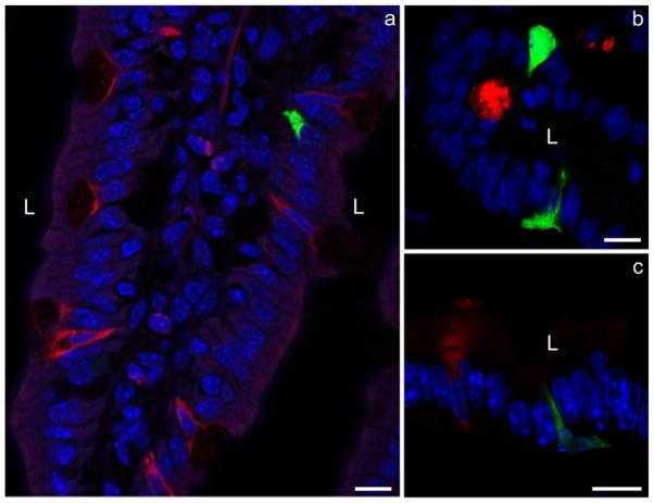
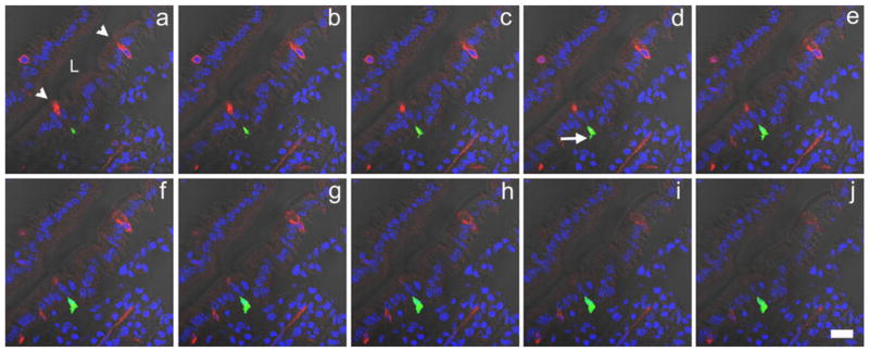
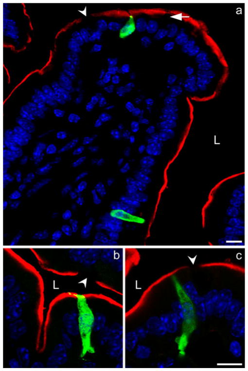
Cholecystokinin (CCK) is secreted by neuroendocrine cells comprising 0.1%-0.5% of the mucosal cells in the upper small intestine. Using CCK promoter-driven green fluorescent protein (GFP) expression in transgenic mice, we have applied immunofluorescence techniques to analyze the morphology of CCK cells. GFP and CCK colocalize in neuroendocrine cells with little aberrant GFP expression. CCK-containing cells are either flask- or spindle-shaped, and in some cells, we have found dendritic processes similar to pseudopods demonstrated for gut somatostatin-containing D cells. Most pseudopods are short, the longest process visualized extending across three cells. Pseudopods usually extend to adjacent cells but some weave between neighboring cells. Dual processes have also been observed. Three-dimensional reconstructions suggest that processes are not unidirectional and thus are unlikely to be involved in migration of CCK cells from the crypt up the villus. Abundant CCK immunostaining is present in the pseudopods, suggesting that they release CCK onto the target cell. In order to identify the type of cells being targeted, we have co-stained sections with antibodies to chromogranin A, trefoil factor-3, and sucrase-isomaltase. CCK cell processes almost exclusively extend to sucrase-isomaltase-positive enterocytes. Thus, CCK cells have cellular processes possibly involved in paracrine secretion.


Figures

Fig. 1
Fig. 1
Colocalization of CCK (cholecystokinin) and GFP (green fluorescent protein) in mouse intestinal CCK cells. a Endogenously fluorescent GFP (green) in a CCK cell. b The same CCK cell as seen in a stained with a rabbit GFP antiserum (red) demonstrating that only cells endogenously expressing GFP stain positively for GFP in the mouse duodenum. c Merged images of a, b. d Endogenously fluorescent GFP in a CCK cell. e The same cell as seen in d stained with a rabbit CCK antibody demonstrating that only CCK cells express GFP in the mouse duodenum. f Merged images of d, e. g CCK cell in which endogenous GFP has been visualized by immunostaining with an anti-GFP serum raised in chickens. h The same CCK cell as seen in g stained with a rabbit CCK antibody demonstrating that GFP and CCK can be immunostained by using primary antisera derived from a different species. i Merged images of g, h. The orientation of the CCK cells relative to the lumen of the intestine and the base of the cells is shown in a and is the same in all other panels (blue DAPI nuclear staining). Bar 5 μm
Fig. 2
Fig. 2
a Two yellow CCK cells at low power exhibiting both endogenous GFP expression (green) and positive immunostaining for GFP with a rabbit anti-GFP serum (red). b Two yellow CCK cells at low power exhibiting both positive immunostaining for CCK with a rabbit anti-CCK serum (red) and for GFP with a chicken anti-GFP serum (green). Note the basal CCK cell processes (arrows). Both photomicrographs were taken with fluorescence (blue DAPI nuclear staining) and differential interference contrast (DIC) optics (L lumen). Bar 20 μm
Fig. 3
Fig. 3
af Examples of CCK-GFP cells stained with chick or rabbit GFP antiserum extending basal cell processes (arrows) to neighboring cells. gi, jl Two examples of colocalization (yellow) of CCK (red) with chick GFP (green) in the basal cell processes of CCK cells. CCK is present in the pseudopods (arrows) together with GFP (blue DAPI nuclear staining). Bar 5 μm
Fig. 4
Fig. 4
Three-dimensional reconstruction of a CCK cell located at the base of a villus (a). The same cell (shown in all panels) possesses a bifurcated pseudopod that extends into spaces other than immediately up or down the crypt-villus axis (bi). GFP is immunostained with chick GFP antibody (green) and CCK with rabbit CCK antibody (red). Both fluorophores are represented as maximum projections (blue DAPI nuclear staining). X-axis, Y-axis, and Z-axis (green, red, and blue, respectively) are shown (bottom left) to demonstrate that each image (bi) is rotated 45° along the Y-axis. The crypt-villus axis is directly opposite the X-axis. Bars 10 μm (a), 5 μm (i)
Fig. 5
Fig. 5
Double-immunofluorescence staining of sections with antibodies against intestinal trefoil factor-3 (red) to identify goblet cells and with either CCK or rabbit GFP antibodies (green). The basal CCK cell processes do not contact the goblet cells positive for intestinal trefoil factor-3 (L lumen, blue DAPI nuclear staining). Bar 10 μm
Fig. 6
Fig. 6
A series of Z-stack images (aj: 0.49 μm apart) of a portion of the mouse duodenum double-stained for GFP (green) in CCK cells and intestinal trefoil factor-3 (red) in goblet cells to demonstrate that CCK basal cell processes are not directed to goblet cells (arrowheads goblet cells positive for intestinal trefoil factor-3, arrow basal CCK cell process, L lumen). Photomicrographs were taken with both fluorescence and DIC optics (blue DAPI nuclear staining). Bar 20 μm
Fig. 7
Fig. 7
Double-immunofluorescence staining of sections with antibodies to sucrase-isomaltase (red) to identify enterocytes and rabbit GFP (green) to identify CCK cells. CCK cells and their basal cell processes are seen to be closely associated with the neighboring enterocytes. Depending on the plane of the section, either no sucrase-isomaltase staining (arrowheads) or faint staining is seen when a goblet cell (arrow) is present (L lumen, blue DAPI nuclear staining). Bar 10 μm

References

    1. Alumets J, Ekelund M, El Munshid HA, Hakanson R, Loren I, Sundler F. Topography of somatostatin cells in the stomach of the rat: possible functional significance. Cell Tissue Res. 1979;202:177–188. - PubMed
    1. Berthoud HR, Patterson LM. Anatomical relationship between vagal afferent fibers and CCK-immunoreactive entero-endocrine cells in the rat small intestinal mucosa. Acta Anat (Basel) 1996;156:123–131. - PubMed
    1. Buchan AM, Polak JM, Solcia E, Capella C, Hudson D, Pearse AG. Electron immunohistochemical evidence for the human intestinal I cell as the source of CCK. Gut. 1978;19:403–407. - PMC - PubMed
    1. Dockray GJ. The versatility of the vagus. Physiol Behav. 2009;97:531–536. - PubMed
    1. Duckworth CA, Pritchard DM. Suppression of apoptosis, crypt hyperplasia, and altered differentiation in the colonic epithelia of bak-null mice. Gastroenterology. 2009;136:943–952. - PubMed

MeSH terms

LinkOut - more resources