. 2010 Oct;67(19):3355-69.
doi: 10.1007/s00018-010-0434-3. Epub 2010 Jun 27.

SAM domain-dependent activity of PfTKL3, an essential tyrosine kinase-like kinase of the human malaria parasite Plasmodium falciparum

Affiliations

SAM domain-dependent activity of PfTKL3, an essential tyrosine kinase-like kinase of the human malaria parasite Plasmodium falciparum

Abdirahman Abdi et al. Cell Mol Life Sci. 2010 Oct.

Abstract

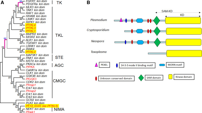
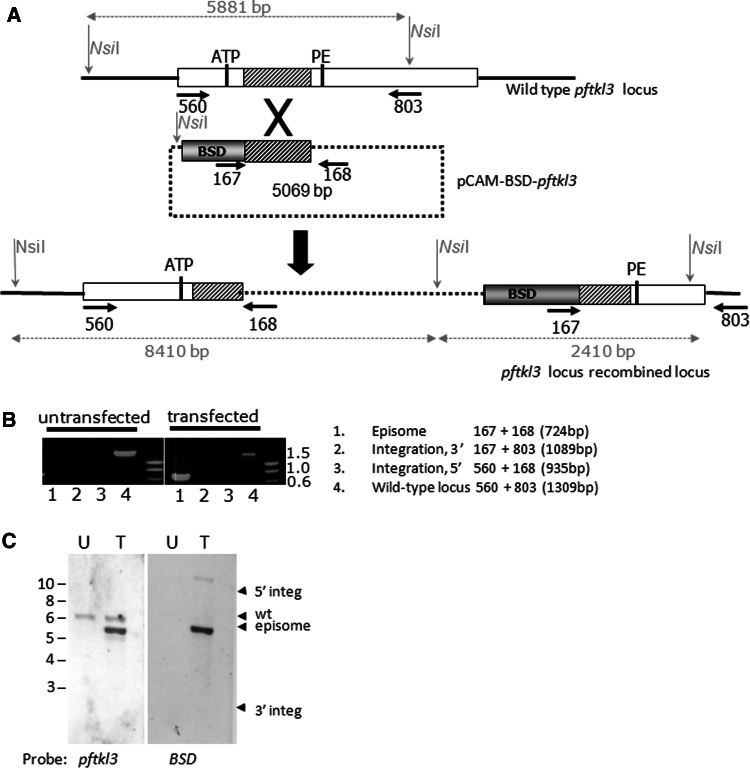
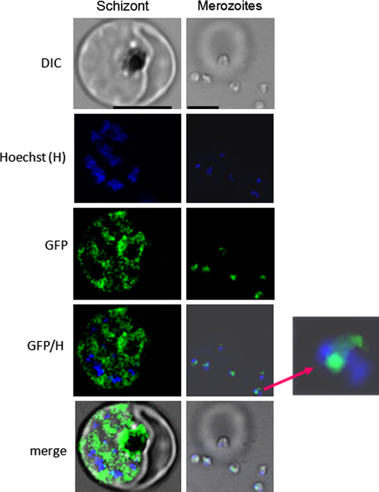
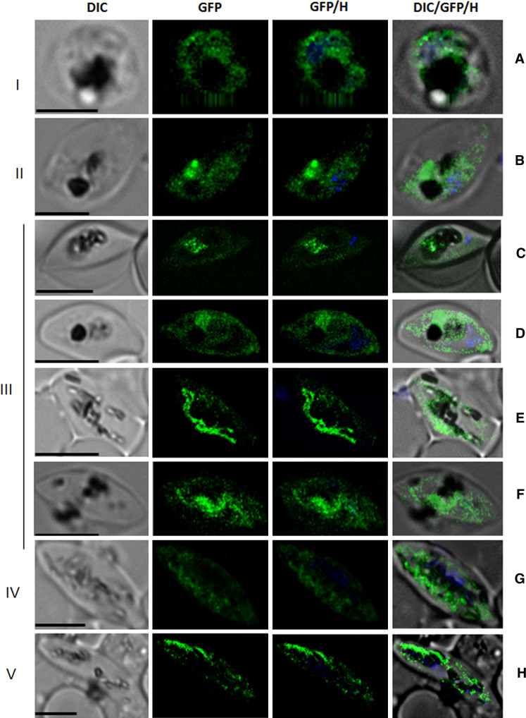
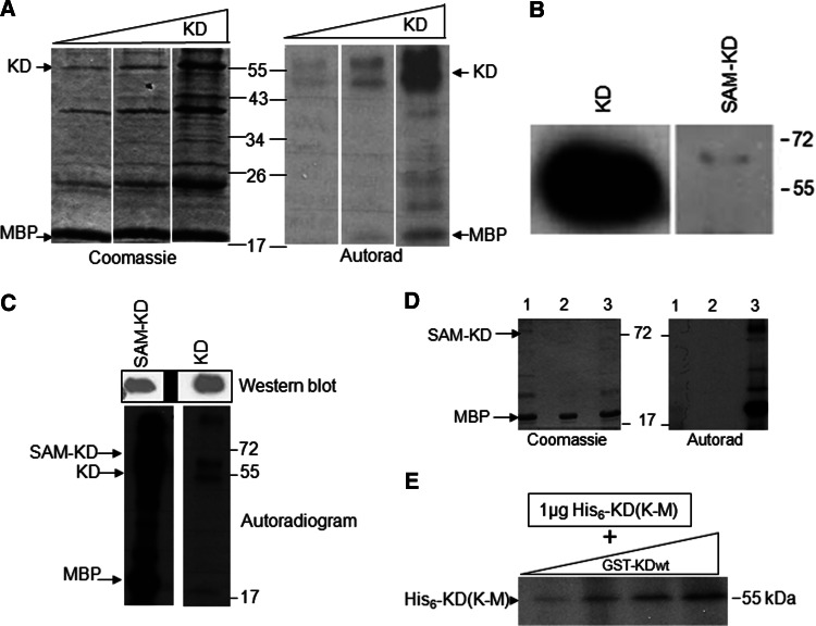
Over the last decade, several protein kinases inhibitors have reached the market for cancer chemotherapy. The kinomes of pathogens represent potentially attractive targets in infectious diseases. The functions of the majority of protein kinases of Plasmodium falciparum, the parasitic protist responsible for the most virulent form of human malaria, remain unknown. Here we present a thorough characterisation of PfTKL3 (PF13_0258), an enzyme that belongs to the tyrosine kinase-like kinase (TKL) group. We demonstrate by reverse genetics that PfTKL3 is essential for asexual parasite proliferation in human erythrocytes. PfTKL3 is expressed in both asexual and gametocytes stages, and in the latter the protein co-localises with cytoskeleton microtubules. Recombinant PfTKL3 displays in vitro autophosphorylation activity and is able to phosphorylate exogenous substrates, and both activities are dramatically dependent on the presence of an N-terminal "sterile alpha-motif" domain. This study identifies PfTKL3 as a validated drug target amenable to high-throughput screening.


Figures

Fig. 1
Fig. 1
Phylogeny and domain organisation of PfTKL3. a Phylogenetic tree of the kinase domain sequences from representatives of the human (black) and P. falciparum (red) kinome families. The five sequences identified as P. falciparum TKLs in Ref. [10] are highlighted in yellow. b Schematic representation of PfTKL3 homologs in Apicomplexa
Fig. 2
Fig. 2
Targeted gene disruption of pftkl3. a Gene disruption strategy. The restriction enzyme (Nsi1) sites used for Southern blot analysis and the sizes of the expected fragments are shown. The arrows (dark bold) indicate the location of primers used in PCR analysis. BSD is a blasticidin-resistance marker. b PCR analysis. The amplicons are diagnostic for: lane 1, episome; lane 2, 3′ boundary of integrated plasmid; lane 3, 5′ boundary of integrated plasmid; lane 4, wild-type locus. The numbers 560, 167, 168 and 803 represent the primers used (sequences in Supplemental Table 1). c Southern blot analysis. Genomic DNA from untransfected (U) and pCAM-BSD-PfTKL3 transfected (T) 3D7 parasites was digested with Nsi1 and probed with PfTKL3 (left panel) and BSD (right panel) probes
Fig. 3
Fig. 3
GFP- and HA-tagging of the pftkl3 locus. a C-terminal tagging strategy. The location of PCR primers used for genotyping is indicated by numbered arrows. See the text for details. b PCR analysis of the GFP tagged locus: Genomic DNA from untransfected and transfected 3D7 parasites was subjected to PCR analysis using the primers numbered in a. The amplicons are diagnostic for: lane 1, episome; lane 2, wild type; lane 3, 3′ boundary of integrated plasmid; lane 4, 5′ boundary of integrated plasmid. The numbers 560, 167, 168, 803 and 806 represent the primers used (sequences in Supplemental Table 1). c PCR analysis of the HA-tagged locus. This was performed as in b, except the DNA was from pCAM-BSD-PfTKL3dHA-transfected parasites, and primer 639 was used instead of primer 806. d Southern blot analysis. Genomic DNA from untransfected (U) and pCAM-BSD-PfTKL3GFP-transfected (T) 3D7 parasites was digested with Nsi1 and probed with a pftkl3 probe. e Western blot analysis. Protein extracts (10 μg/lane) from dHA-tagged (lane 1) and GFP-tagged (lane 2) lines were separated by SDS-PAGE, transferred to nitrocellulose and probed with anti-GFP (left), anti-HA (right) or anti-PfGR (bottom, loading control) antibodies
Fig. 4
Fig. 4
Visualisation of GFP-tagged PfTKL3 in asexual parasites. Live asexual cultures from the GFP-tagged line were observed by fluorescence microscopy. A signal was detected only in the schizont and free merozoites. A typical image is shown for both these stages. A 4× relative enlargement of a merozoite is shown to the right. Scale bar: 5 μm
Fig. 5
Fig. 5
Visualisation of GFP-tagged PfTKL3 in gametocytes. A live gametocyte from the PfTKL-GFP line was observed by fluorescence microscopy. Signals were observed throughout gametocyte development. See text for details. Scale bar: 5 μm
Fig. 6
Fig. 6
Co-localisation studies. a An antibody against the parasitophorous vacuole membrane protein Pfs16 was used to counterstain PfTKL3-GFP expressing gametocytes. b An anti-tubulin antibody (anti-TubA) was used to counterstain PfTKL3-GFP expressing gametocytes. The white arrows indicate where the GFP and TubA signal concentrate in late stages of gametocytes. See text for details. Scale bar: 5 μm
Fig. 7
Fig. 7
Kinase activity of recombinant PfTKL3. a Kinase activity of recombinant His6-PfTKL3 kinase domain (KD); 0.5, 1 or 4 μg of His6-PfTKL3-KD wild type was used in a kinase reaction containing 5 μg of myelin basic protein (MBP). Left panel: Coomassie stain of the gel; right panel: autoradiograph. b Western blot of soluble PfTKL3 KD and PfTKL3 SAM-KD recovered from 400 ml of bacterial culture, using an anti-His6 antibody. Equal volumes were loaded, corresponding to 40 ml of culture. c MBP kinase activity assay using equal amounts of SAM-KD and KD forms of recombinant PfTKL3. Top panel: Western blot using an anti-His6 antibody showing equal loading of both forms of the recombinant enzyme. Bottom panel: autoradiogram. d Kinase dead-mutant controls. Kinase assays were performed with the wild-type recombinant SAM-KD PfTKL3 (lane 3), and with the K → M (lane 1) or D → N (lane 2) kinase-dead mutants using MBP as substrate. Left panel: Coomassie staining of the gel; right panel: autoradiogram. e Autophosphorylation in trans: 1 μg of the catalytically inactive His6-tagged (K → M) mutant kinase domain was used as a substrate in kinase assay using increasing amounts of PfTKL3-KD (twofold serial dilutions from the highest point, which contained 6 μg of active wild-type GST-PfTKL3 KD)
Fig. 8
Fig. 8
Mutagenesis of the PfTKL3 SAM domain. a Model for the stimulation of kinase activity by an adjacent SAM domain. Adapted from [23]. b Alignment of the SAM domains of ETV6 and PfTKL3. The five helices in the secondary structure of ETV6 SAM domain are shown as grey cylinders. The ETV6 residues and corresponding PfTKL3 that occur in the interacting interfaces called mid-loop (ML) H3 and end-helix (EH) H5 are shown in red. Numbering is from the N-terminal boundary of each SAM domain. c Effect of SAM domain mutations on the recovery of soluble recombinant SAM-KD PfTKL3. Top panel: Western blot (anti-His6) of wild-type, V54E and H35E recovered from 400 ml of bacterial cultures. Bottom panel: The V54E mutation results in a yield that is sufficient for detection by Coomassie staining
Fig. 9
Fig. 9
MBP-SAM domain pull down assay. Purified GST-SAM was incubated with MBP-SAM (lane 1) or MBP alone (lane 2). GST-SAM was incubated with the lysis buffer as negative control (lane 3). Amylose-agarose beads were added to the mixture, incubated and washed. The bound proteins were eluted and subjected to SDS-PAGE and Western blot analysis. The reciprocal experiment (GST pull-down assay) and a gel showing the purified proteins used in this experiment can be found in Supplemental Fig. S4

Similar articles

Cited by

References

    1. White NJ, Nosten F, Looareesuwan S, Watkins WM, Marsh K, Snow RW, Kokwaro G, Ouma J, Hien TT, Molyneux ME, Taylor TE, Newbold CI, Ruebush TK, 2nd, Danis M, Greenwood BM, Anderson RM, Olliaro P. Averting a malaria disaster. Lancet. 1999;353:1965–1967. doi: 10.1016/S0140-6736(98)07367-X. - DOI - PubMed
    1. Snow RW, Trape JF, Marsh K. The past, present and future of childhood malaria mortality in Africa. Trends Parasitol. 2001;17:593–597. doi: 10.1016/S1471-4922(01)02031-1. - DOI - PubMed
    1. Dondorp AM, Nosten F, Yi P, Das D, Phyo AP, Tarning J, Lwin KM, Ariey F, Hanpithakpong W, Lee SJ, Ringwald P, Silamut K, Imwong M, Chotivanich K, Lim P, Herdman T, An SS, Yeung S, Singhasivanon P, Day NP, Lindegardh N, Socheat D, White NJ. Artemisinin resistance in plasmodium falciparum malaria. N Engl J Med. 2009;361:455–467. doi: 10.1056/NEJMoa0808859. - DOI - PMC - PubMed
    1. Dondorp AM, Yeung S, White L, Nguon C, Day NP, Socheat D, von Seidlein L. Artemisinin resistance: current status and scenarios for containment. Nat Rev Microbiol 8:272–280 - PubMed
    1. Hanks SK. Genomic analysis of the eukaryotic protein kinase superfamily: a perspective. Genome Biol. 2003;4:111. doi: 10.1186/gb-2003-4-5-111. - DOI - PMC - PubMed

Publication types

LinkOut - more resources