

Inhibition of angiopoietin-2 in LuCaP 23.1 prostate cancer tumors decreases tumor growth and viability
- PMID: 20583134
- PMCID: PMC3104406
- DOI: 10.1002/pros.21216
Inhibition of angiopoietin-2 in LuCaP 23.1 prostate cancer tumors decreases tumor growth and viability
Abstract
Background: Angiopoietin-2 is expressed in prostate cancer (PCa) bone, liver, and lymph node metastases, whereas, its competitor angiopoietin-1 has limited expression in these tissues. Therefore, we hypothesized that the inhibition of angiopoietin-2 activity in PCa will impede angiogenesis, tumor growth, and alter bone response in vivo.
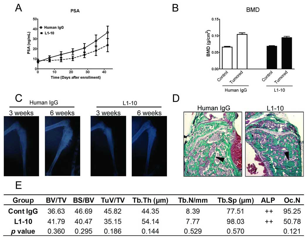
Methods: To test our hypothesis we used L1-10, a peptide-Fc fusion that inhibits interactions between angiopoietin-2 and its receptor tie2. We blocked angiopoietin-2 activity using L1-10 in established subcutaneous and intra-tibial LuCaP 23.1 xenografts. We then determined the effect of L1-10 on survival, tumor growth, serum PSA, proliferation, microvessel density, and angiogenesis-associated gene expression in subcutaneous tumors. We also determined serum PSA, tumor area, and bone response in intra-tibial tumors.
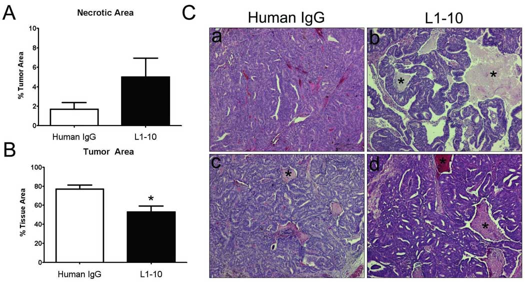
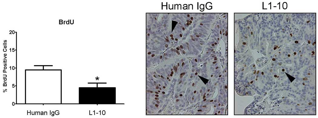
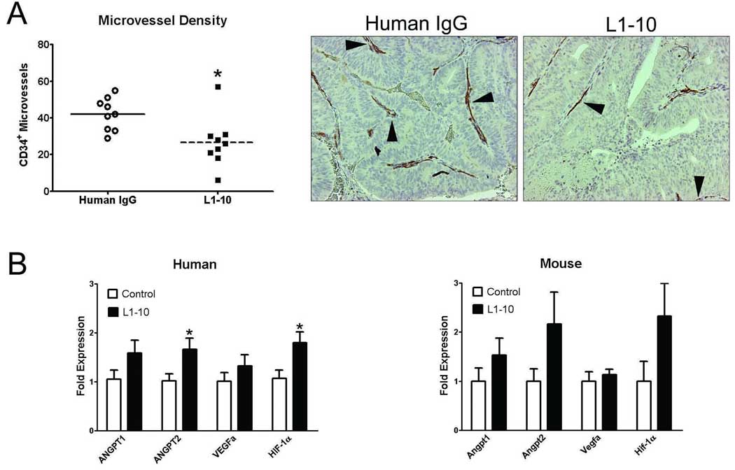
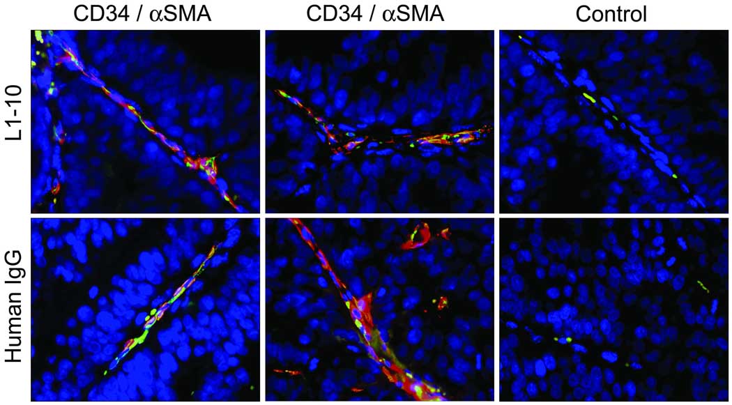
Results: The administration of L1-10 decreased tumor volume and serum PSA, and increased survival in SCID mice bearing subcutaneous LuCaP 23.1 tumors. Histomorphometric analysis, showed a further significant decrease in tumor epithelial area within the L1-10 treated LuCaP 23.1 subcutaneous tumors (P=0.0063). There was also a significant decrease in cell proliferation (P=0.012), microvessel density (P=0.012), and a significant increase in ANGPT-2 and HIF-1α mRNA expression (P≤0.05) associated with L1-10 treatment. Alternatively, in LuCaP 23.1 intra-tibial tumors L1-10 treatment did not significantly change serum PSA, tumor area or bone response.
Conclusions: Our results demonstrate that inhibiting angiopoietin-2 activity impedes angiogenesis and growth of LuCaP 23.1 PCa xenografts. Based on these data, we hypothesize that angiopoietin-2 inhibition in combination with other therapies may represent a potential therapy for patients with metastatic disease.
© 2010 Wiley-Liss, Inc.
Figures

Similar articles
-
The effect of osteoprotegerin administration on the intra-tibial growth of the osteoblastic LuCaP 23.1 prostate cancer xenograft.Clin Exp Metastasis. 2004;21(5):381-7. doi: 10.1007/s10585-004-2869-0. Clin Exp Metastasis. 2004. PMID: 15672862
-
Inhibition of ERG Activity in Patient-derived Prostate Cancer Xenografts by YK-4-279.Anticancer Res. 2017 Jul;37(7):3385-3396. doi: 10.21873/anticanres.11705. Anticancer Res. 2017. PMID: 28668826
-
Characterization of a novel androgen-sensitive, prostate-specific antigen-producing prostatic carcinoma xenograft: LuCaP 23.Clin Cancer Res. 1996 Jun;2(6):1039-48. Clin Cancer Res. 1996. PMID: 9816265
-
Establishing human prostate cancer cell xenografts in bone: induction of osteoblastic reaction by prostate-specific antigen-producing tumors in athymic and SCID/bg mice using LNCaP and lineage-derived metastatic sublines.Int J Cancer. 1998 Sep 11;77(6):887-94. doi: 10.1002/(sici)1097-0215(19980911)77:6<887::aid-ijc15>3.0.co;2-z. Int J Cancer. 1998. PMID: 9714059
-
Targeted chemotherapy with cytotoxic bombesin analogue AN-215 inhibits growth of experimental human prostate cancers.Int J Cancer. 2006 Jan 1;118(1):222-9. doi: 10.1002/ijc.21292. Int J Cancer. 2006. PMID: 16003723
Cited by
-
Animal Models and Their Role in Imaging-Assisted Co-Clinical Trials.Tomography. 2023 Mar 16;9(2):657-680. doi: 10.3390/tomography9020053. Tomography. 2023. PMID: 36961012 Free PMC article. Review.
-
Spheroid culture of LuCaP 136 patient-derived xenograft enables versatile preclinical models of prostate cancer.Clin Exp Metastasis. 2016 Apr;33(4):325-37. doi: 10.1007/s10585-016-9781-2. Epub 2016 Feb 12. Clin Exp Metastasis. 2016. PMID: 26873136
-
Serum biomarker analysis in patients with recurrent spontaneous abortion.Mol Med Rep. 2017 Sep;16(3):2367-2378. doi: 10.3892/mmr.2017.6890. Epub 2017 Jun 30. Mol Med Rep. 2017. PMID: 28677727 Free PMC article.
-
The natural compound atraric acid suppresses androgen-regulated neo-angiogenesis of castration-resistant prostate cancer through angiopoietin 2.Oncogene. 2022 Jun;41(23):3263-3277. doi: 10.1038/s41388-022-02333-7. Epub 2022 May 5. Oncogene. 2022. PMID: 35513564 Free PMC article.
-
Establishment and serial passage of cell cultures derived from LuCaP xenografts.Prostate. 2013 Sep;73(12):1251-62. doi: 10.1002/pros.22610. Epub 2013 Jun 6. Prostate. 2013. PMID: 23740600 Free PMC article.
References
-
- Folkman J, Greenspan HP. Influence of geometry on control of cell growth. Biochim Biophys Acta. 1975;417:211–236. - PubMed
-
- Folkman J. Tumor angiogenesis: a possible control point in tumor growth. Ann Intern Med. 1975;82:96–100. - PubMed
-
- Yancopoulos GD, Davis S, Gale NW, Rudge JS, Wiegand SJ, Holash J. Vascular-specific growth factors and blood vessel formation. Nature. 2000;407:242–248. - PubMed
-
- Ferrer FA, Miller LJ, Andrawis RI, Kurtzman SH, Albertsen PC, Laudone VP, Kreutzer DL. Angiogenesis and prostate cancer: in vivo and in vitro expression of angiogenesis factors by prostate cancer cells. Urology. 1998;51:161–167. - PubMed
Publication types
MeSH terms
Substances
Grants and funding
LinkOut - more resources
Full Text Sources
Other Literature Sources
Medical
Research Materials
Miscellaneous
