

The mechanical function of the periodontal ligament in the macaque mandible: a validation and sensitivity study using finite element analysis
- PMID: 20584094
- PMCID: PMC3039782
- DOI: 10.1111/j.1469-7580.2010.01257.x
The mechanical function of the periodontal ligament in the macaque mandible: a validation and sensitivity study using finite element analysis
Retraction in
-
Retraction. ‘The mechanical function of the periodontal ligament in the macaque mandible: a validation and sensitivity study using finite element analysis’ by O. Panagiotopoulou, K. Kupczik and S.N. Cobb.J Anat. 2015 May;226(5):498. doi: 10.1111/joa.12322. J Anat. 2015. PMID: 26176031 Free PMC article. No abstract available.
Expression of concern in
-
Expression of concern.J Anat. 2014 Apr;224(4):527. doi: 10.1111/joa.12149. J Anat. 2014. Retraction in: J Anat. 2015 May;226(5):498. doi: 10.1111/joa.12322. PMID: 24754054 Free PMC article. Retracted. No abstract available.
Abstract
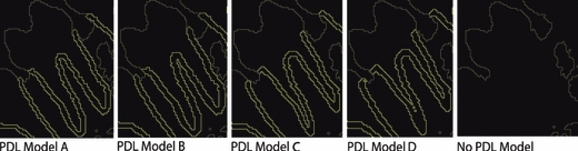
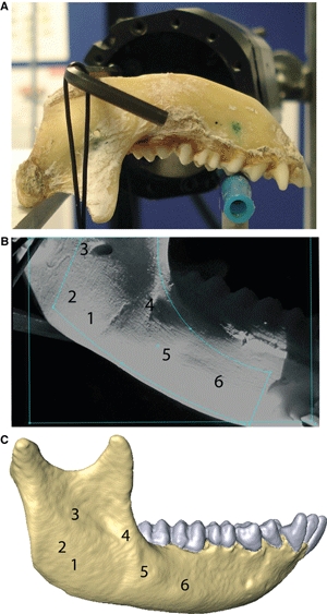
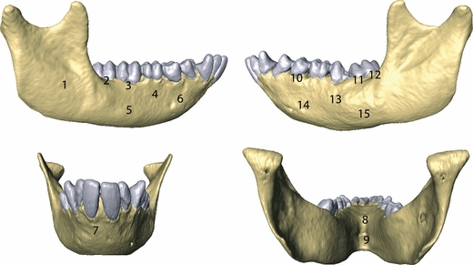
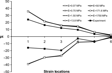
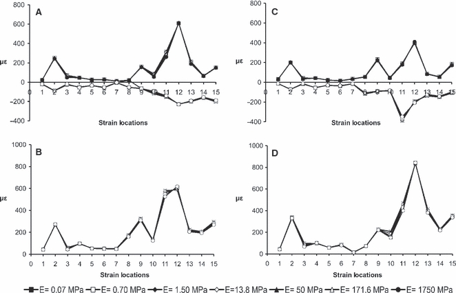
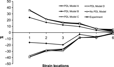
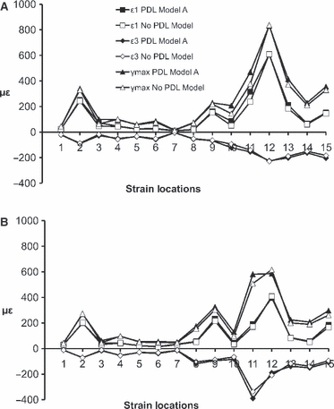
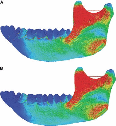
Whilst the periodontal ligament (PDL) acts as an attachment tissue between bone and tooth, hypotheses regarding the role of the PDL as a hydrodynamic damping mechanism during intraoral food processing have highlighted its potential importance in finite element (FE) analysis. Although experimental and constitutive models have correlated the mechanical function of the PDL tissue with its anisotropic, heterogeneous, viscoelastic and non-linear elastic nature, in many FE simulations the PDL is either present or absent, and when present is variably modelled. In addition, the small space the PDL occupies and the inability to visualize the PDL tissue using μCT scans poses issues during FE model construction and so protocols for the PDL thickness also vary. In this paper we initially test and validate the sensitivity of an FE model of a macaque mandible to variations in the Young's modulus and the thickness of the PDL tissue. We then tested the validity of the FE models by carrying out experimental strain measurements on the same mandible in the laboratory using laser speckle interferometry. These strain measurements matched the FE predictions very closely, providing confidence that material properties and PDL thickness were suitably defined. The FE strain results across the mandible are generally insensitive to the absence and variably modelled PDL tissue. Differences are only found in the alveolar region adjacent to the socket of the loaded tooth. The results indicate that the effect of the PDL on strain distribution and/or absorption is restricted locally to the alveolar bone surrounding the teeth and does not affect other regions of the mandible.
© 2010 The Authors. Journal of Anatomy © 2010 Anatomical Society of Great Britain and Ireland.
Figures
References
-
- Andersen KL, Pedersen EH, Melsen B. Material parameters and stress profiles within the periodontal ligament. Am J Orthod Dentofacial Orthop. 1991;99:427–440. - PubMed
-
- Atmaram HF, Mohammed H. Estimation of physiologic stresses with a natural tooth considering fibrous PDL structure. J Dent Res. 1981;60:873–877. - PubMed
-
- Berkovitz BKB, Moxham BJ, Newman HN. The Periodontal Ligament in Health and Disease. London: Mosby-Wolfe; 1995.
-
- Bien SM. Fluid dynamic mechanisms which regulate tooth movement. In: Staple PH, editor. Advances in Oral Biology. Vol. 2. London: Academic Press; 1966. pp. 173–201. - PubMed
-
- Byers MR. Sensory innervation of periodontal ligament of rat molars consists of unencapsulated Ruffini-like mechanoreceptors and free nerve endings. J Comp Neurol. 1985;231:500–518. - PubMed
Publication types
MeSH terms
LinkOut - more resources
Full Text Sources
