. 2010 Jun 28:10:20.
doi: 10.1186/1475-2867-10-20.

New clinically relevant, orthotopic mouse models of human chondrosarcoma with spontaneous metastasis

Affiliations

New clinically relevant, orthotopic mouse models of human chondrosarcoma with spontaneous metastasis

Jonathan Cm Clark et al. Cancer Cell Int. .

Abstract

Background: Chondrosarcoma responds poorly to adjuvant therapy and new, clinically relevant animal models are required to test targeted therapy.

Methods: Two human chondrosarcoma cell lines, JJ012 and FS090, were evaluated for proliferation, colony formation, invasion, angiogenesis and osteoclastogenesis. Cell lines were also investigated for VEGF, MMP-2, MMP-9, and RECK expression. JJ012 and FS090 were injected separately into the mouse tibia intramedullary canal or tibial periosteum. Animal limbs were measured, and x-rayed for evidence of tumour take and progression. Tibias and lungs were harvested to determine the presence of tumour and lung metastases.

Results: JJ012 demonstrated significantly higher proliferative capacity, invasion, and colony formation in collagen I gel. JJ012 conditioned medium stimulated endothelial tube formation and osteoclastogenesis with a greater potency than FS090 conditioned medium, perhaps related to the effects of VEGF and MMP-9. In vivo, tumours formed in intratibial and periosteal groups injected with JJ012, however no mice injected with FS090 developed tumours. JJ012 periosteal tumours grew to 3 times the non-injected limb size by 7 weeks, whereas intratibial injected limbs required 10 weeks to achieve a similar tumour size. Sectioned tumour tissue demonstrated features of grade III chondrosarcoma. All JJ012 periosteal tumours (5/5) resulted in lung micro-metastases, while only 2/4 JJ012 intratibial tumours demonstrated metastases.

Conclusions: The established JJ012 models replicate the site, morphology, and many behavioural characteristics of human chondrosarcoma. Local tumour invasion of bone and spontaneous lung metastasis offer valuable assessment tools to test the potential of novel agents for future chondrosarcoma therapy.


Figures

Figure 1
Figure 1
Chondrosarcoma cell morphology and proliferation. a) JJ012 demonstrated high pleomorphism and nuclear atypia with prominent nucleoli. b) FS090 had more distinct polarity and less nuclear atypia. c) JJ012 proliferated at a higher rate than FS090 (*p < 0.05). d) Prominent colony formation was noted for JJ012 suspended in collagen I gel. e) FS090 did not form colonies after 5 days.
Figure 2
Figure 2
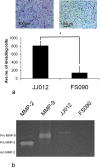
Chondrosarcoma cell invasion. a) A higher number of JJ012 cells invaded Matrigel compared with FS090 (* p < 0.05). b) Greater invasive capacity corresponded with the presence of pro MMP-9 in JJ012 conditioned media on gelatin zymography. This was in contrast with undetectable pro MMP-9 in FS090. No MMP-2 was detected for either cell line and no activated forms were present.
Figure 3
Figure 3
Chondrosarcoma angiogenesis. a) Conditioned media from JJ012 induced a higher number of tube formations compared with FS090 (* p < 0.05). b) Greater angiogenic capacity in JJ012 corresponded with the presence of VEGF on western blotting, in comparison with virtually undetectable VEGF in FS090. The anti-angiogenic protein RECK was prominently expressed by FS090, but not by JJ012.
Figure 4
Figure 4
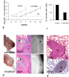
In vivo growth and progression of chondrosarcoma cell lines. a) This graph plots the injected to non-injected limb volume ratios for the study groups at each time point. Periosteal injected JJ012 developed into tumours most rapidly, followed 2-3 weeks later by intratibial injected JJ012. FS090 did not form tumours in either implantation site. b) Periosteal tumours expanded one area of the limb while intratibial tumours grew in a circumferential manner. On histology both tumours (T) invaded cortex, but leaving the epiphysis (E) largely intact. On radiographs, lytic regions (black arrow), and sclerotic (grey arrow) were noted in both models. c) 5/5 animals injected with periosteal JJ012 demonstrated lung metastases, in contrast with 2/4 animals in the intratibial group. d) Lung metastases (M) were lobular in appearance, and were positive on Alizarin red staining (arrows), indicating calcification.
Figure 5
Figure 5
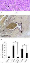
Tumour histology and osteoclast involvement. a) Pleomorphic, ovoid cells (*) with an additional spindle cell population (arrows) were noted for JJ012 tumours. b) Dedifferentiated chondrosarcoma from a human de novo tumour demonstrated some similar ovoid cells without a spindle cell component. c) In periosteal and intratibial tumours, TRAP staining (arrows) revealed prominent osteoclasts at the interface between tumour (T) and bone cortex (C), and this was associated with bone matrix erosion and a mass of dead cellular material (D). d) An osteoclastogenesis assay demonstrated higher osteoclasts formed by JJ012 media (CM) compared with FS090 media (CM) (* p < 0.05), and the same relationship for chondrosarcoma and mouse monocyte (RAW 246.7) co-culture (* p < 0.05).

References

    1. Björnsson J, McLeod RA, Unni KK, Ilstrup DM, Pritchard DJ. Primary chondrosarcoma of long bones and limb girdles. Cancer. 1998;83:2105–2119. doi: 10.1002/(SICI)1097-0142(19981115)83:10<2105::AID-CNCR9>3.0.CO;2-U. - DOI - PubMed
    1. Lee FY, Mankin HJ, Fondren G, Gebhardt MC, Springfield DS, Rosenberg AE, Jennings LC. Chondrosarcoma of bone: an assessment of outcome. J Bone Joint Surg Am. 1999;81:326–338. doi: 10.1302/0301-620X.81B5.9588. - DOI - PubMed
    1. Bruns J, Elbracht M, Niggemeyer O. Chondrosarcoma of bone: an oncological and functional follow-up study. Ann Oncol. 2001;12:859–864. doi: 10.1023/A:1011162118869. - DOI - PubMed
    1. Pring ME, Weber KL, Unni KK, Sim FH. Chondrosarcoma of the pelvis. A review of sixty-four cases. J Bone Joint Surg Am. 2001;83-A:1630–1642. - PubMed
    1. Johnson S, Tetu B, Ayala AG, Chawla SP. Chondrosarcoma with additional mesenchymal component (dedifferentiated chondrosarcoma). I. A clinicopathologic study of 26 cases. Cancer. 1986;58:278–286. doi: 10.1002/1097-0142(19860715)58:2<278::AID-CNCR2820580213>3.0.CO;2-6. - DOI - PubMed

LinkOut - more resources