. 2010 Jun 28:5:18.
doi: 10.1186/1749-7922-5-18.

Angiography and embolisation for solid abdominal organ injury in adults - a current perspective

Affiliations

Angiography and embolisation for solid abdominal organ injury in adults - a current perspective

Adam Wallis et al. World J Emerg Surg. .

Abstract

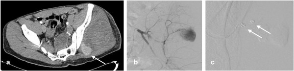
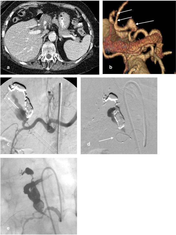
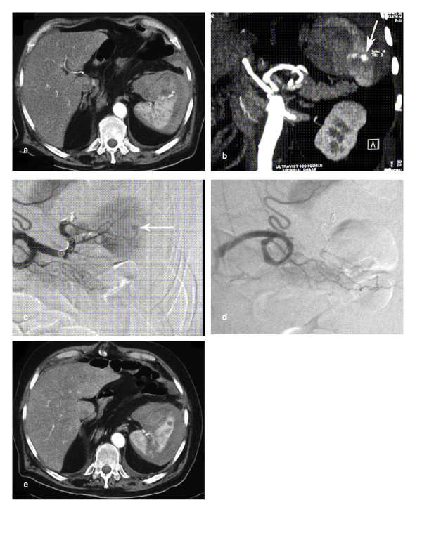
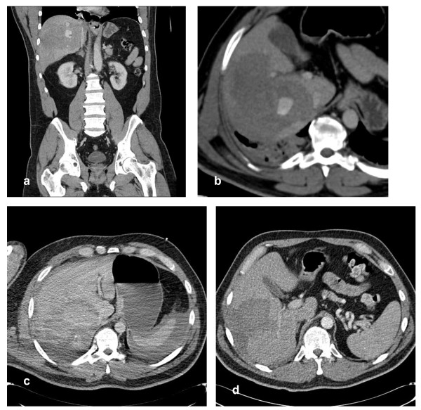
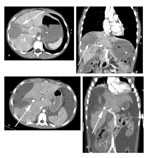
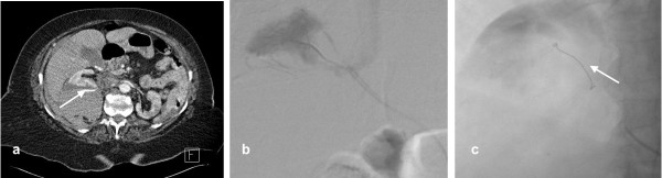
Over the past twenty years there has been a shift towards non-operative management (NOM) for haemodynamically stable patients with abdominal trauma. Embolisation can achieve haemostasis and salvage organs without the morbidity of surgery, and the development and refinement of embolisation techniques has widened the indications for NOM in the management of solid organ injury. Advances in computed tomography (CT) technology allow faster scanning times with improved image quality. These improvements mean that whilst surgery is still usually recommended for patients with penetrating injuries, multiple bleeding sites or haemodynamic instability, the indications for NOM are expanding.We present a current perspective on angiography and embolisation in adults with blunt and penetrating abdominal trauma with illustrative examples from our practice including technical advice.


Figures

Figure 1
Figure 1
a) Axial arterial phase contrast enhanced CT in a 23 year old man following a stab wound to the left buttock demonstrates haematoma within the gluteus muscles. Contrast enhancement medially (arrow) represents active haemorrhage from the superior gluteal artery (Somatom sensation, 24 slice,Siemens, Erlangen, Germany). b) A Cobra catheter was negotiated into the posterior (somatic) left internal iliac artery from an ipsilateral approach. Active haemorrhage from a branch of the superior gluteal artery was demonstrated. c) A microcatheter system (Progreat) was negotiated into the bleeding vessel and 2 microcoils (Boston Scientific vortex fibred) were deployed (arrows). This completely abolished the bleeding with good perfusion of the buttock post procedure.
Figure 2
Figure 2
a) A patient with vasculitic hepatic artery aneurysms presented following minor trauma. Axial contrast enhanced CT demonstrates haematoma around a pseudoaneurysm (arrow) indicating that this is the likely cause of recent haemodynamic instability. b) 3D volume rendered reconstruction demonstrates 3 aneurysms arising from a branch of the left hepatic artery (arrows). The right hepatic artery arose from the SMA. c) Selective arteriogram of the coeliac axis with standard catheter after 2 aneurysms had been embolised with onyx (ev3, Irvine, CA, USA). The cast of the onyx is demonstrated, and some distal embolisation (arrow) of onyx. d) A microcatheter is demonstrated within the final bleeding aneurysm (arrow). e) A selective angiogram demonstrates onyx filling all aneurysms and maintained patency of the gastroduodenal artery.
Figure 3
Figure 3
a) Axial CT of a 73 year old man with iatrogenic splenic injury following chest drain insertion. An active bleeding point in the spleen (arrow) with surrounding haematoma was demonstrated. b) Coronal CT reconstruction showing a tortuous splenic artery and bleeding point (arrow). These allowed optimal catheter choice for arteriography. c) A Tracker-18 microcatheter system with a Fasdasher 0.014 in wire (Boston Scientific, Maple Grove, MN, USA) were used to achieve access distally within the splenic circulation. After several unsuccessful attempts at superselective catheterisation of the branch supplying the bleeding point, 4 platinum Vortex-18 diamond-shaped coils (Boston Scientific) were deployed sequentially in the main splenic artery distal to the dorsal pancreatic branch. 2 initial coils migrated past the required branch and there is ongoing bleeding from the spleen (arrow). d) The next 2 coils achieved occlusion of the main splenic artery with preservation of branches to the dorsal pancreas and upper pole of the spleen. e) Axial CT at 1 week showed a small splenic infarct where the initial coils had migrated distally. Arterial supply to the spleen was preserved with some flow through the main splenic artery coils.
Figure 4
Figure 4
a) Coronal contrast enhanced arterial phase CT reconstruction showing contrast blush in a contained right lobe haematoma due to blunt inury. b) Axial CT demonstrates the blush. c) Scan at 18 hours showing no blush but capsular rupture with intraperitoneal blood. d) Follow up CT at 9 weeks showing resolving right lobe haematoma.
Figure 5
Figure 5
a) Axial contrast enhanced CT of a teenager who sustained a handlebar injury to the abdomen. Large laceration/haematoma (arrow) and no active extravasation. b) Coronal reconstruction demonstrates free fluid around the right lobe of the liver (arrow) and the extent of the laceration. He was managed conservatively initially but deteriorated several days later. c) An emergency CT showed a contrast blush (arrow). d) Maximimum intensity projections demonstrated that the most likely cause was the right anterior portal vein (arrow). At operation (not by our team) biliary peritonitis was found but there was no active bleeding and subsequent hepatic angiography was negative.
Figure 6
Figure 6
a) A 76 year old lady on warfarin presented with abdominal and back pain following a fall. Contrast enhanced axial CT demonstrates retroperitoneal haematoma associated with a ruptured right kidney and evidence of active contrast extravasaion (arrow). b) Selective catheterisation of the right kidney showed a bleeding focus in the upper pole. c) The branch to the upper pole was selectively catheterised and embolised using a single platinum coil (arrow). Post procedure renal arteriogram demonstrated cessation of haemorrhage.

Similar articles

Cited by

References

    1. World Health Organisation. Guidelines for essential trauma care. 2004. http://whqlibdoc.who.int/publications/2004/9241546409.pdf
    1. Deunk J, Brink M, Dekker HM. Predictors for the selection of patients for abdominal CT after blunt trauma: a proposal for a diagnostic algorithm. Ann Surg. 2010;251(3):512–520. doi: 10.1097/SLA.0b013e3181cfd342. - DOI - PubMed
    1. Fang JF, Wong YC, Lin BC. Usefulness of multidetector computed tomography for the initial assessment of blunt abdominal trauma patients. World J Surg. 2006;30:176–182. doi: 10.1007/s00268-005-0194-7. - DOI - PubMed
    1. Zealley IA, Chakraverty S. The role of interventional radiology in trauma. BMJ. 2010;340:c497. doi: 10.1136/bmj.c497. - DOI - PubMed
    1. Hilbert P, zur Nieden K, Hofmann GO. New aspects in the emergency room management of critically injured patients A multislice CT-orientated care algorithm. Injury. 2007;38:552–558. doi: 10.1016/j.injury.2006.12.023. - DOI - PubMed

LinkOut - more resources