Rupture of a large vertebral artery aneurysm following proximal occlusion
- PMID: 20584435
- PMCID: PMC3403788
- DOI: 10.1177/159101990501100108
Rupture of a large vertebral artery aneurysm following proximal occlusion
Abstract
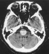
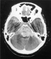
Proximal occlusion of the vertebral artery is regarded as a safe and effective method of treating aneurysms of the vertebral artery or the vertebrobasilar junction unsuitable for treatment by neck clipping. Complications known to develop after this procedure include ischemic lesions of the perforators and other areas. There are only a limited number of reports on early rupture of aneurysm following proximal occlusion of the vertebral artery for the treatment of unruptured aneurysm. We recently encountered a case of large aneurysm of the vertebral artery identified after detection of brainstem compression. This patient underwent proximal occlusion of the vertebral artery with a coil and developed a fatal rupture of the aneurysm ten days after proximal occlusion. The patient was a 72-year-old woman who had complained of dysphagia and unsteadiness for several years. An approximately 20 mm diameter aneurysm was detected in her left vertebral artery. She underwent endovascular treatment, that is, her left vertebral artery was occluded with coils at a point proximal to the aneurysm. Her initial post-procedure course was uneventful. However, she suddenly developed right-side hemiparesis nine days after procedure. At that time, CT scan suggested sudden thrombosis of the aneurysm. Right vertebral angiography revealed a small part of the aneurysm. She was treated conservatively. Ten days after the procedure, she suffered massive subarachnoid haemorrhage. Both the present case and past reports suggest that proximal occlusion of the vertebral artery is effective in treating relatively large aneurysms unsuitable for treatment by neck clipping or trapping. However, when the bifurcation of the posterior inferior cerebellar artery (PICA) is distal to the occluded point in cases where the PICA bifurcates from the aneurysm or the neck region, blood supply to the aneurysm may persist because anterograde blood flow to the PICA may be preserved. Therefore, clinicians must consider the possibility of aneurysm rupture after proximal occlusion in the following cases: 1) when the aneurysm is large or giant, but non-thrombosed; 2) when thrombosis occurs soon after the procedure; 3) when postoperative angiography shows partial filling of the aneurysm with contrast agent through the contralateral vertebral artery of basilar artery or the cervical muscle branches.
Figures


Similar articles
-
Outflow Occlusion with Occipital Artery-Posterior Inferior Cerebellar Artery Bypass for Growing Vertebral Artery Fusiform Aneurysm with Ischemic Onset: A Case Report.J Stroke Cerebrovasc Dis. 2015 Aug;24(8):e223-6. doi: 10.1016/j.jstrokecerebrovasdis.2015.04.020. Epub 2015 Jun 5. J Stroke Cerebrovasc Dis. 2015. PMID: 25979424
-
[Intraaneurysmal embolization and parent artery trapping to treat a giant partial thrombosed vertebral artery aneurysm after surgical proximal clipping].No Shinkei Geka. 2000 Sep;28(9):817-22. No Shinkei Geka. 2000. PMID: 11025883 Japanese.
-
[Giant partially thrombosed aneurysm of the vertebral artery: a case report and literature review].Zh Vopr Neirokhir Im N N Burdenko. 2016;80(5):106-115. doi: 10.17116/neiro2016805106-115. Zh Vopr Neirokhir Im N N Burdenko. 2016. PMID: 28635695 Review. Russian.
-
[Endovascular management and classification of the dissecting aneurysms of the vertebral artery].Zhonghua Yi Xue Za Zhi. 2017 Jun 20;97(23):1773-1777. doi: 10.3760/cma.j.issn.0376-2491.2017.23.004. Zhonghua Yi Xue Za Zhi. 2017. PMID: 28647997 Chinese.
-
[A case of de novo aneurysm of the distal posterior inferior cerebellar artery with intraventricular hemorrhage].No Shinkei Geka. 1996 May;24(5):469-73. No Shinkei Geka. 1996. PMID: 8692375 Review. Japanese.
Cited by
-
Therapeutic dilemmas regarding giant aneurysms of the intracranial vertebral artery causing medulla oblongata compression.Neuroradiol J. 2022 Apr;35(2):137-151. doi: 10.1177/19714009211042881. Epub 2021 Sep 3. Neuroradiol J. 2022. PMID: 34477003 Free PMC article.
-
A case of lateral medullary infarction after endovascular trapping of the vertebral artery dissecting aneurysm.J Korean Neurosurg Soc. 2012 Mar;51(3):160-3. doi: 10.3340/jkns.2012.51.3.160. Epub 2012 Mar 31. J Korean Neurosurg Soc. 2012. PMID: 22639714 Free PMC article.
-
Microsurgical partial trapping for the treatment of unclippable vertebral artery aneurysms: Experience from 27 patients and review of literature.World Neurosurg X. 2023 Dec 5;21:100256. doi: 10.1016/j.wnsx.2023.100256. eCollection 2024 Jan. World Neurosurg X. 2023. PMID: 38163051 Free PMC article.
-
Intracranial Fusiform and Circumferential Aneurysms of the Main Trunk: Therapeutic Dilemmas and Prospects.Front Neurol. 2021 Jul 9;12:679134. doi: 10.3389/fneur.2021.679134. eCollection 2021. Front Neurol. 2021. PMID: 34305790 Free PMC article. Review.
References
-
- Shintani A, Zervas NT. Consequence of ligation of the vertebral artery. J Neurosurg. 1972;36:447–450. - PubMed
-
- Drake CG. Ligation of the vertebral (unilateral or bilateral) or basilar artery in the treatment of large intracranial aneurysms. J. Neurosurg. 1975;43:255–274. - PubMed
-
- Steinberg GK, Drake CG, Peerless SJ. Deliberate basilar or vertebral artery occlusion in the treatment of intracranial aneurysms. Immediate results and long-term outcome in 201 patients. J Neurosurg. 1993;79:161–173. - PubMed
-
- Kawamata T, Tanikawa T, et al. Rebleeding of intracranial dissecting aneurysm in the vertebral artery following proximal clipping. Neurol Res. 1994;16:141–144. - PubMed
-
- Kitanaka C, Morimoto T, et al. Rebleeding from vertebral artery dissection after proximal clipping. J Neurosurg. 1992;77:466–468. - PubMed
LinkOut - more resources
Full Text Sources
Miscellaneous

