

Vertebroplasty in the treatment of spine disease
- PMID: 20584443
- PMCID: PMC3399747
- DOI: 10.1177/159101990501100403
Vertebroplasty in the treatment of spine disease
Abstract
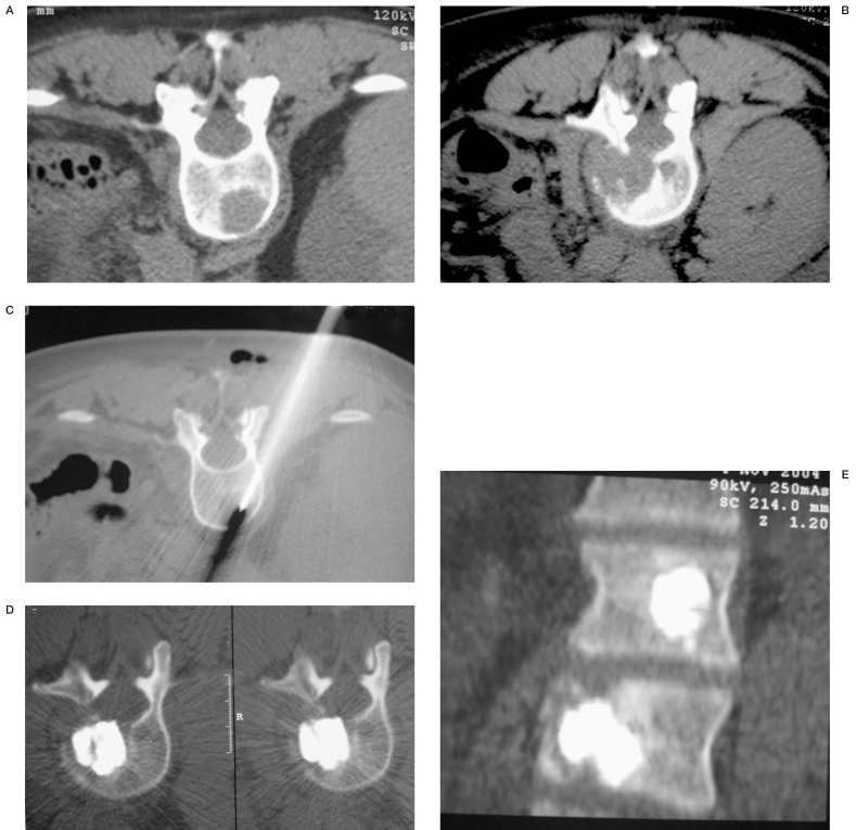
We report our experience in the treatment of thoracic and lumbosacral spinal pain due to vertebral bone fractures. This pathology can be related to osteoporosis but also to metastatic disease and less frequently vertebral haemangioma. From April 2001 through December 2004 we treated 238 patients for a total of 455 vertebral bodies. 175 patients had osteoporosis, 70 had metastasis and 13 had vertebral haemangioma. Sacroplasty was performed in six patients to obtain a cement filling of sacral metastasis. The procedures were mostly performed under fluoroscopy and only in cases of metastasis or sacroplasty was CT/fluoroscopy guidance preferred for optimal filling of the area of osteolysis. We evaluated the results at six and 18 months follow-up and analysed the incidence of new vertebral fractures, vascular and disk leakage and the incidence of major and minor complications. Biopsy was performed only in doubtful cases. We obtained different results considering the etiology of the disease. We obtained a 92% success rate at six months follow-up and 89% success at 18 months follow-up in osteoporosis, a 77% and 72% success rate at six and 18 months follow-up in metastastic patients, and no change at six and 18 months follow-up in patients with vertebral haemangioma in which the success rate was of 95%. We noted extravertebral leakage in 41% of vertebral bodies of which 31% were treated at the level of the vascular space and only 10% at the level of the disk space, and symptomatic in only two cases (acute compressive radiculitis, medically treated and resolved within a month). Six patients presented new fractures in the adjacent vertebral body and 30% had a partial recovery in the height of the vertebral body with kyphosis curve reduction. Vertebroplasty is a good technique to obtain spine pain relief and has a low incidence of side effects. Good quality equipment is important to obtain these results.
Figures


Similar articles
-
Vertebroplasty in the treatment of back pain.Radiol Med. 2005 Mar;109(3):208-19. Radiol Med. 2005. PMID: 15775889 English, Italian.
-
An in vivo comparison of the potential for extravertebral cement leak after vertebroplasty and kyphoplasty.Spine (Phila Pa 1976). 2002 Oct 1;27(19):2173-8; discussion 2178-9. doi: 10.1097/00007632-200210010-00018. Spine (Phila Pa 1976). 2002. PMID: 12394934 Clinical Trial.
-
Vertebroplasty: cement leakage into the disc increases the risk of new fracture of adjacent vertebral body.AJNR Am J Neuroradiol. 2004 Feb;25(2):175-80. AJNR Am J Neuroradiol. 2004. PMID: 14970015 Free PMC article.
-
Repeated percutaneous vertebroplasty for refracture of cemented vertebrae.Arch Orthop Trauma Surg. 2011 Jul;131(7):927-33. doi: 10.1007/s00402-010-1236-7. Epub 2010 Dec 30. Arch Orthop Trauma Surg. 2011. PMID: 21191607
-
Comparison of Percutaneous Vertebroplasty and Balloon Kyphoplasty for the Treatment of Single Level Vertebral Compression Fractures: A Meta-analysis of the Literature.Pain Physician. 2015 May-Jun;18(3):209-22. Pain Physician. 2015. PMID: 26000665 Review.
Cited by
-
Management of vertebral re-fractures after vertebroplasty in osteoporotic patients.Interv Neuroradiol. 2009 Jul 29;15(2):153-7. doi: 10.1177/159101990901500203. Epub 2009 Sep 1. Interv Neuroradiol. 2009. PMID: 20465892 Free PMC article.
-
Percutaneous sacroplasty for the management of painful pathologic fracture in a multiple myeloma patient: Case report and review of the literature.Neuroradiol J. 2017 Feb;30(1):80-83. doi: 10.1177/1971400916678642. Epub 2016 Nov 25. Neuroradiol J. 2017. PMID: 27888274 Free PMC article. Review.
-
Percutaneous treatment of a sacral metastasis with combined embolization, cryoablation, alcohol ablation and sacroplasty for local tumor and pain control.Interv Neuroradiol. 2013 Jun;19(2):250-3. doi: 10.1177/159101991301900217. Epub 2013 May 21. Interv Neuroradiol. 2013. PMID: 23693052 Free PMC article.
-
Vertebroplasty and kyphoplasty: friends or foes?Radiol Med. 2008 Dec;113(8):1171-84. doi: 10.1007/s11547-008-0301-6. Epub 2008 Oct 3. Radiol Med. 2008. PMID: 18836816 English, Italian.
-
Update of vertebral cementoplasty in porotic patients.Interv Neuroradiol. 2015 Jun;21(3):372-80. doi: 10.1177/1591019915582364. Epub 2015 May 26. Interv Neuroradiol. 2015. PMID: 26015527 Free PMC article. Review.
References
-
- Amar AP, Larsen DW, et al. Percutaneous transpedicular polymethylmethacrylate vertebroplasty for the treatment of spinal compression fractures. Neurosurgery. 2001;49:1105–1114. - PubMed
-
- Andreula C, Muto M, Leonardi M. Interventional spinal procedures. Eur J Radiol. 2004;50:112–119. - PubMed
-
- Anita A, Uppin MD, et al. Occurrence of new vertebral body fracture after percutaneous vertebroplasty in patients with osteoporosis. Radiology. 2003;226:119–124. - PubMed
-
- Bajaj S, Saag KG. Osteoporosis: evaluation and treatment. Curr Womens Health Rep. 2003;3:418–124. - PubMed
LinkOut - more resources
Full Text Sources
Miscellaneous
