

The anastomotic venous circle of the base of the brain
- PMID: 20584444
- PMCID: PMC3399748
- DOI: 10.1177/159101990501100404
The anastomotic venous circle of the base of the brain
Abstract
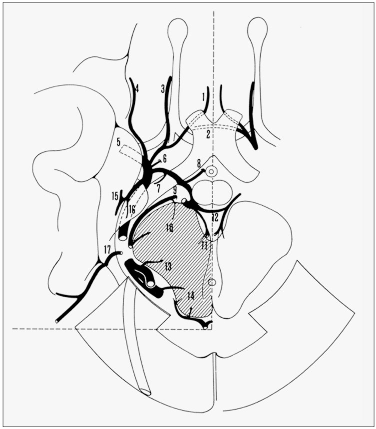
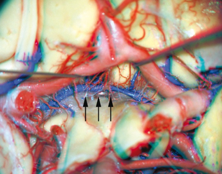
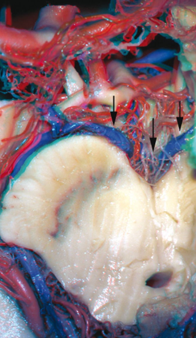
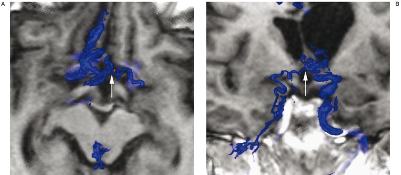
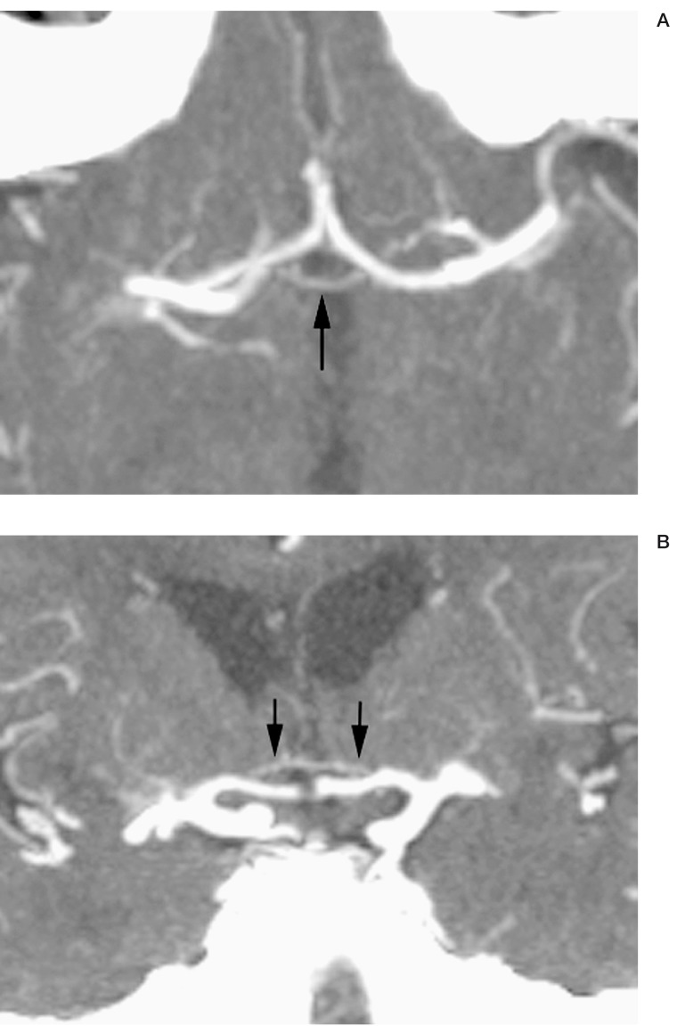
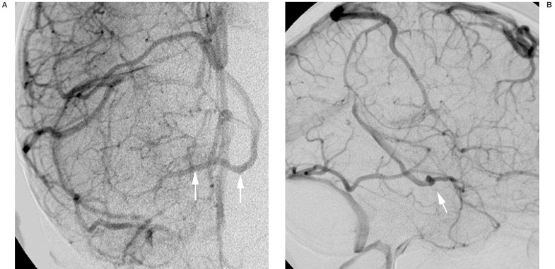
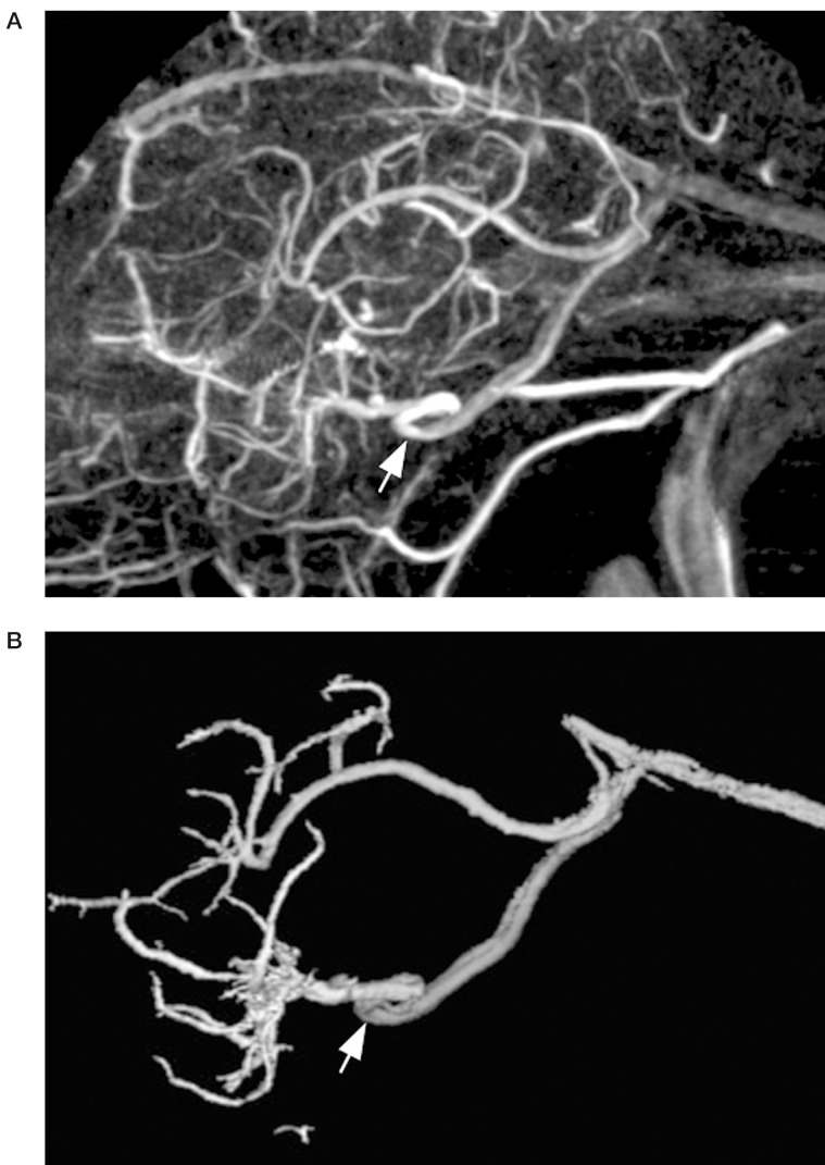
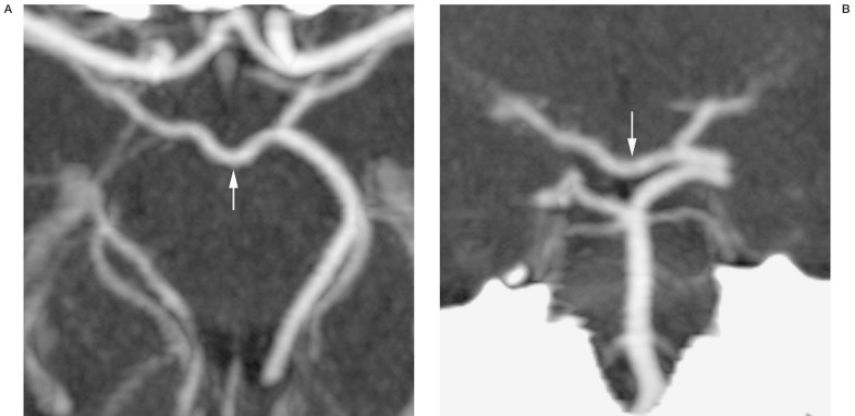
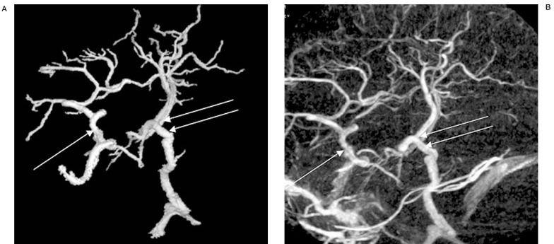
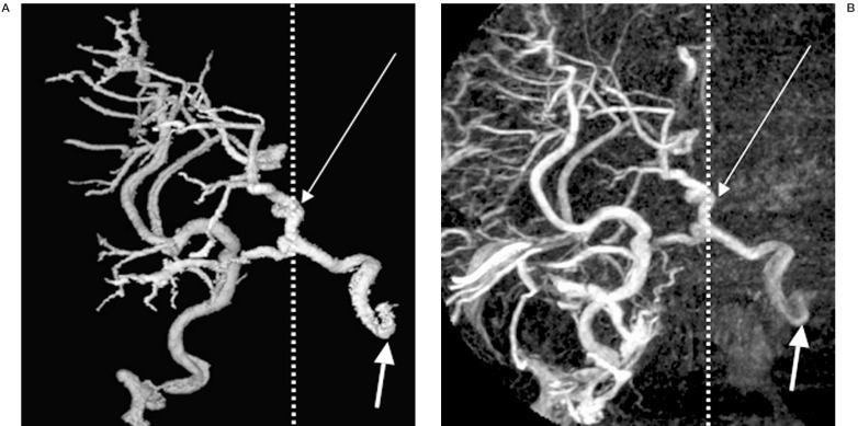
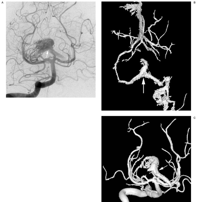
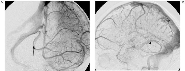
Adjacent to the arterial circle of Willis at the base of the brain, there is an anastomotic circle of veins linking the right and left halves of the cerebral deep venous system. This venous circle is formed by anterior and posterior transverse anastomotic channels (the anterior and posterior communicating veins), and paramedian longitudinal vessels (the basal veins of Rosenthal). This collateral venous network has received considerably less attention than its arterial counterpart, but is its functional homologue. Although inconstant, it can be seen readily with current neuroimaging techniques including three-dimensional digital subtraction venographic phase 3D arteriography (3D-DSV) and CT venography (CTV). The venous circle represents a route of contralateral venous drainage that may become important, particularly when segments of the basal vein are absent (with or without complex DVA), or in high flow states including arteriovenous shunts that access the deep venous system.We review the gross anatomy and provide examples of the radiologic imaging of this venous circle.
Figures
Similar articles
-
The venous circle of Trolard.Bratisl Lek Listy. 2008;109(4):180-1. Bratisl Lek Listy. 2008. PMID: 18814436
-
The Venous Circle of Trolard: An Anatomical Study with Application to Approaches to the Basal Brain.World Neurosurg. 2023 Jul;175:e238-e242. doi: 10.1016/j.wneu.2023.03.059. Epub 2023 Mar 20. World Neurosurg. 2023. PMID: 36940805
-
Transverse anastomoses of the veins at the base of the brain.Neuroradiology. 1976;12(3):165-9. doi: 10.1007/BF00341861. Neuroradiology. 1976. PMID: 1004702
-
Venous collateral circulation of the extracranial cerebrospinal outflow routes.Curr Neurovasc Res. 2009 Aug;6(3):204-12. doi: 10.2174/156720209788970054. Epub 2009 Aug 1. Curr Neurovasc Res. 2009. PMID: 19534716 Review.
-
Bone subtraction 3D CT venography for the evaluation of cerebral veins and venous sinuses: imaging techniques, normal variations, and pathologic findings.AJR Am J Roentgenol. 2014 Feb;202(2):W169-75. doi: 10.2214/AJR.13.10985. AJR Am J Roentgenol. 2014. PMID: 24450700 Review.
Cited by
-
Contralateral extensive cerebral hemorrhagic venous infarction caused by retrograde venous reflux into the opposite basal vein of Rosenthal in posttraumatic carotid-cavernous fistula: A case report and literature review.Interv Neuroradiol. 2018 Oct;24(5):546-558. doi: 10.1177/1591019918776615. Epub 2018 May 19. Interv Neuroradiol. 2018. PMID: 29781369 Free PMC article. Review.
-
Scanning fast and slow: current limitations of 3 Tesla functional MRI and future potential.Front Phys. 2014 Feb 11;2:00001. doi: 10.3389/fphy.2014.00001. Front Phys. 2014. PMID: 28164083 Free PMC article.
-
Atypical telencephalic reflux pattern in a cavernous sinus dural AV fistula related to an anatomical variation of the basal vein of Rosenthal.Interv Neuroradiol. 2024 Jun 7:15910199241260758. doi: 10.1177/15910199241260758. Online ahead of print. Interv Neuroradiol. 2024. PMID: 38847128 Free PMC article.
-
Mapping the circle of Trolard: A holistic view of its role in neurosurgical practice, anatomical perspectives and clinical applications.Brain Spine. 2024 Mar 23;4:102789. doi: 10.1016/j.bas.2024.102789. eCollection 2024. Brain Spine. 2024. PMID: 38562441 Free PMC article. Review.
-
Transdural Segment of the Radicular Vein in Spinal Dural Arteriovenous Fistula.Neurointervention. 2017 Mar;12(1):57-58. doi: 10.5469/neuroint.2017.12.1.57. Epub 2017 Mar 6. Neurointervention. 2017. PMID: 28316872 Free PMC article. No abstract available.
References
-
- Duvernoy H. The superficial veins of the human brain. Berlin, Heidelberg, New York: Springer; 1975.
-
- Rosenthal F. De intimis cerebri venis seu de venae magnae galena ramis. Nova acta physico-medica Academie Caesareae Leopoldino-Carolinae, Naturae cuiosorum. 1824;12:302–312.
-
- Padget DH. The cranial venous system in man in reference to development, adult configuration and relation to the arteries. Am J Anat. 1956;98:307–355. - PubMed
-
- Padget DH. The development of the cranial venous system in man, from the viewpoint of comparative anatomy. Contributions to Embryology. 1957;36:79–140.
-
- Braun JP, Tournade A, Ammerich H. Transverse anastomoses of the veins at the base of the brain. Neuroradiology. 1956;12:165–169. - PubMed
LinkOut - more resources
Full Text Sources
