. 2005 Oct 5;11(Suppl 1):73-80.
doi: 10.1177/15910199050110S112. Epub 2005 Oct 27.

Endovascular Treatment of AVMs in Glasgow

Affiliations

Endovascular Treatment of AVMs in Glasgow

J J Bhattacharya et al. Interv Neuroradiol. .
No abstract available


Figures

Figure 1
Figure 1
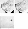
27-year-old woman who presented with seizures. MRI revealed a small occipital AVM related to the tentorium cerebelli. A) Pre-embolization angiogram shows single feeder. B) Microcatheter (Magic 1.2) navigated with Mirage wire into nidus. A single injection of Glubran-2 sealed the AVM. C) Follow-up angiography at 1 year confirms cure.
Figure 2
Figure 2
59-year-old woman with history of seizures and brain haematoma in adolescence. A) Angiography shows left MCA AVM (>3 cm). B) Embolization with NBCA glue via Spinnaker-1.5 (Mizzen-12 wire). Asymptomatic venous penetration of glue (arrow). C) After 3 embolization sessions.
Figure 3
Figure 3
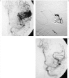
A 42-year-old woman with severe seizures poorly controlled (4-5 seizures per week) on maximal drug therapy. A) MRI shows large incurable AVM. B) Vertebral angiogram shows large occipito-temporal deep AVM and giant basilar aneurysm. C) After 3 embolization sessions nidal flow markedly reduced. Seizure frequency reduced to one in three months.
Figure 4
Figure 4
A 24-year-old woman with severe migrainous headaches and a proliferative angiopathy pattern AVM. A) T2-weighted MRI demonstrates AVM nidus interspersed with brain parenchyma. Functional MRI confirmed functioning brain tissue within nidus. B, C) Angiography demonstrates large diffuse nidus.
Figure 5
Figure 5
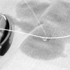
A Spinnaker-1.8 microcatheter being flushed with saline solution prior to use. The proximal leak is readily visualised.

References

    1. Al-Shahi R, Bhattacharya JJ, et al. Prospective, population-based detection of intracranial vascular malformations in adults: The Scottish Intracranial Vascular Malformation Study (SIVMS) Stroke. 2003;34:1163–1169. - PubMed
    1. Al-Shahi R, Bhattacharya JJ, et al. Scottish Intracranial Vascular Malformation Study (SIVMS): Evaluation of methods, ICD-10 coding, and potential sources of bias in a prospective, population-based cohort. Stroke. 2003;34:1156–1162. - PubMed
    1. Bhattacharya JJ, Thammaroj J. Vein of Galen malformations. J Neurol Neurosurg Psych. 2003;74:42–44. - PMC - PubMed
    1. Thammaroj J, Bhattacharya JJ. Interventional neuroradiology in neonates and children. Radiology Now. 2002;19:811.
    1. Al-Shahi R, Pal N, et al. Observer agreement in the angiographic assessment of arteriovenous malformations of the brain. Stroke. 2002;33:1501–1508. - PubMed

LinkOut - more resources