

Anatomic variations of the deep cerebral veins,tributaries of Basal vein of rosenthal: embryologic aspects of the regressed embryonic tentorial sinus
- PMID: 20584491
- PMCID: PMC3399712
- DOI: 10.1177/159101990501100202
Anatomic variations of the deep cerebral veins,tributaries of Basal vein of rosenthal: embryologic aspects of the regressed embryonic tentorial sinus
Abstract
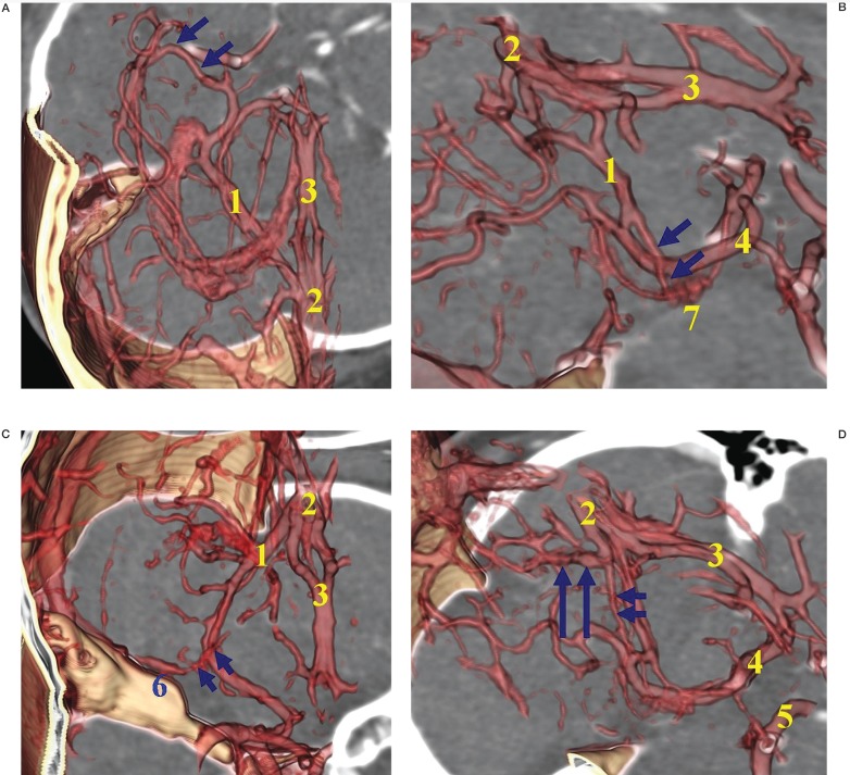
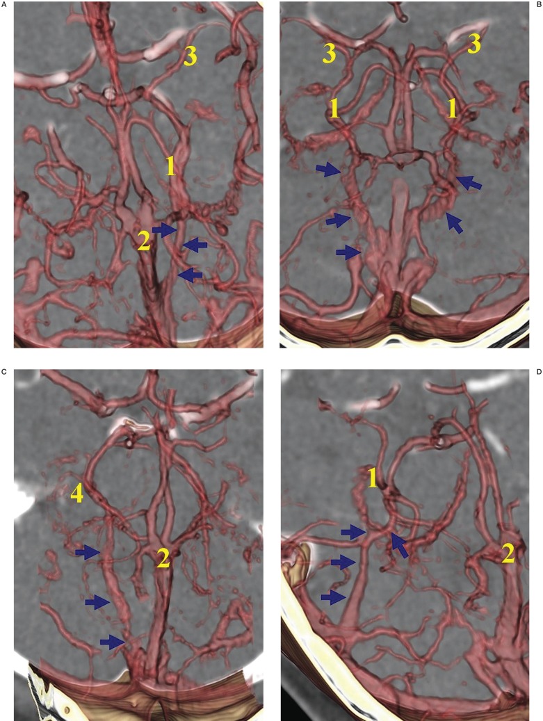
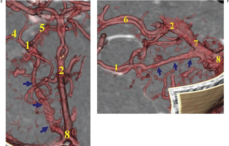
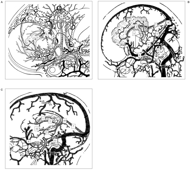
The embryonic tentorial sinus regresses at the 60-80 mm embryologic stage and most of the deep venous channels constitute the basal vein of Rosenthal (BVR). Persisting remnants of the embryonic tentorial sinus can be seen in the adult configuration of the BVR.We tried to explain the anatomic representations of the BVR associated with the remnant embryonic tentorial sinus. A total 41 patients and 82 hemispheres were included in this study. CT angiography was performed in all patients as screening for cerebrovascular disease or other intracranial disorders. A separate workstation and 3D software were used to evaluate the cranial deep venous systems with 3D volume rendering techniques, thin-slice MIP images, and MPR techniques for the analysis of complicated angioarchitecture. Variations of the BVR were classified according to the developmental alterations of efferent pathways into four groups: telencephalic group (A) including tributaries of the uncal vein, inferior frontal vein, anterior communicating vein, and inferior striatal vein; diencephalic group (B) of the interior ventricular vein and peduncular vein; tegmental bridging group (C) of the longitudinal LMV anastomosis; tectal group (D) of the superior vermian vein and internal occipital vein in relation to the Galenic connection. The BVR constituted from the embryonic tentorial sinus was also assessed and the developmental aspects reviewed. Remnant embryonic tentorial sinus was visualized in 12% (10/82) of hemispheres, all of them invariably connected with the telencephalic (A) and diencephalic (B) groups. Most of those connections (9/10) to basal venous tributaries originated from the medial tentorial sinus except one case from the lateral tentorial sinus. No Galenic connections of the BVR were identified in 10% (8/82). Various tributaries of the BVR were classified as: Telencephalic group (A) 43% (35/82), Diencephalic group (B) 35% (29/82), Bridging group (C) 11% (9/82), and Tectal group (D) 6% (5/82). Four cases (5%) were unclassified and revealed only small basal tributaries of the BVR without connection to the great vein of Galen. Anatomic variations of the BVR connected with persistent embryonic tentorial sinus could often be demonstrated in adult configurations considering the embryologic aspects of developmental regression and secondary cerebral venous adaptations.
Figures
References
-
- Lasjaunias P, Berenstein A, ter Brugge KG. Clinical Vascular Anatomy and Variations. 1 2nd edition. Berlin: Springer-Verlag; 2001. Intracranial venous system. Surgical Neuroangiography.
-
- Wolf BS, Huang YP, Newman CM. The lateral anastomotic mesencephalic vein and other variations in drainage of the basal cerebral vein. Am J Roentgenol Radium Ther Nucl Med. 1963;89:411–422. - PubMed
-
- terbrugge K, Lasjaunias P. Tentorial sinus. Radiologic and anatomic features of a case. Surg Radiol Anat. 1988;10:243–246. - PubMed
-
- San Millan Ruiz D, Fasel JH, et al. Bilateral tentorial sinus drainage of the basal vein (of Rosenthal) Clin Anat. 2003;16:264–268. - PubMed
-
- Casey SO, Alberico RA, et al. Cerebral CT venography. Radiology. 1996;198:163–170. - PubMed
LinkOut - more resources
Full Text Sources
