Comparison of corneal changes after phacoemulsification using BSS Plus versus Lactated Ringer's irrigating solution: a prospective randomised trial
- PMID: 20584708
- PMCID: PMC3061049
- DOI: 10.1136/bjo.2009.172502
Comparison of corneal changes after phacoemulsification using BSS Plus versus Lactated Ringer's irrigating solution: a prospective randomised trial
Abstract
Background/aims: To compare two intraocular irrigating solutions, Balanced Salt Solution Plus (BSS Plus) versus Lactated Ringer's (Ringer), for the preservation of corneal integrity after phacoemulsification.
Methods: 110 patients undergoing phacoemulsification were randomised to either BSS Plus (n=55) or Ringer (n=55) as the irrigating solution. Patients were examined at baseline and at 1, 8, 15, 30 and 60 days postoperatively. Evaluations included specular microscopy to evaluate endothelial cell density (ECD) and endothelial cell size variability (CV), and corneal pachymetry for central corneal thickness (CCT) measurement.
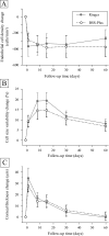
Results: Groups were well balanced regarding baseline ECD, CV and CCT (p>0.05). There was no statistically significant difference between ECD reduction in group BSS Plus 13.1 ± 2.0% and Ringer 9.2 ± 1.9% (p<0.05) at day 60 or in any study visit. There was no statistically significant difference between CV increase in group BSS Plus 23.0 ± 3.0% and Ringer 20.2 ± 4.0% (p<0.05) at day 60 or in any study visit. CCT was significantly increased (p<0.05) at 1, 8, 15 and 30 days postoperatively, returning to baseline at 60 days in both groups. There was no significant difference in CCT increase in both groups at any visit. Interestingly, there were statistically significant correlations between ECD loss and phacoemulsification time (p<0.0001) and ECD loss and irrigation solution volume (p<0.0001) in the Ringer group, but not in the BSS Plus group.
Conclusions: Ringers solution was similar to BSS Plus for corneal preservation in atraumatic cataract surgery. However, our study demonstrates that there is a trend towards lower postoperative endothelial cell density for surgeries with longer phacoemulsification time and higher irrigation volumes if Ringer is used. Trial registration number NCT00801358.
Conflict of interest statement
Figures


Similar articles
-
Effect of irrigating solution and irrigation temperature on the cornea and pupil during phacoemulsification.J Cataract Refract Surg. 2000 Mar;26(3):392-7. doi: 10.1016/s0886-3350(99)00470-8. J Cataract Refract Surg. 2000. PMID: 10713235 Clinical Trial.
-
Effect on corneal endothelial cell loss during phacoemulsification: fortified balanced salt solution versus Ringer lactate.J Cataract Refract Surg. 2012 Sep;38(9):1552-8. doi: 10.1016/j.jcrs.2012.04.036. Epub 2012 Jul 24. J Cataract Refract Surg. 2012. PMID: 22832531 Clinical Trial.
-
Comparison between Ringer's lactate and balanced salt solution on postoperative outcomes after phacoemulsfication: a randomized clinical trial.Indian J Ophthalmol. 2009 May-Jun;57(3):191-5. doi: 10.4103/0301-4738.49392. Indian J Ophthalmol. 2009. PMID: 19384012 Free PMC article. Clinical Trial.
-
Intraocular irrigating solutions. A randomized clinical trial of balanced salt solution plus and dextrose bicarbonate lactated Ringer's solution.Ophthalmology. 1995 Feb;102(2):291-6. doi: 10.1016/s0161-6420(95)31026-3. Ophthalmology. 1995. PMID: 7862416 Clinical Trial.
-
Corneal endothelial cells and acoustic cavitation in phacoemulsification.World J Clin Cases. 2023 Mar 16;11(8):1712-1718. doi: 10.12998/wjcc.v11.i8.1712. World J Clin Cases. 2023. PMID: 36969995 Free PMC article. Review.
Cited by
-
Randomized controlled trial on the safety of intracameral cephalosporins in cataract surgery.Clin Ophthalmol. 2010 Dec 8;4:1499-504. doi: 10.2147/OPTH.S15602. Clin Ophthalmol. 2010. PMID: 21191447 Free PMC article.
-
Narrative review after post-hoc trial analysis of factors that predict corneal endothelial cell loss after phacoemulsification: Tips for improving cataract surgery research.PLoS One. 2024 Mar 21;19(3):e0298795. doi: 10.1371/journal.pone.0298795. eCollection 2024. PLoS One. 2024. PMID: 38512953 Free PMC article. Review.
-
Evaluation of corneal endothelial cell loss following vitrectomy with different endotamponades.Oman J Ophthalmol. 2025 Jun 24;18(2):150-154. doi: 10.4103/ojo.ojo_258_24. eCollection 2025 May-Aug. Oman J Ophthalmol. 2025. PMID: 40666788 Free PMC article.
-
Effect of Nondominant Left-Handed Phacoemulsification Surgery on Corneal Endothelium.Cureus. 2023 Mar 27;15(3):e36744. doi: 10.7759/cureus.36744. eCollection 2023 Mar. Cureus. 2023. PMID: 37123767 Free PMC article.
-
Long-term follow-up of changes in corneal endothelium after primary and secondary intraocular lens implantations in children.Graefes Arch Clin Exp Ophthalmol. 2012 Jun;250(6):925-30. doi: 10.1007/s00417-011-1872-9. Epub 2011 Dec 6. Graefes Arch Clin Exp Ophthalmol. 2012. PMID: 22143676
References
-
- Mishima S. Clinical investigations on the corneal endothelium. Ophthalmology 1982;89:525–30 - PubMed
-
- Bourne WM, Nelson LR, Hodge DO. Central corneal endothelial cell changes over a ten-year period. Invest Ophthalmol Vis Sci 1997;38:779–82 - PubMed
-
- Dick HB, Kohnen T, Jacobi FK, et al. Long-term endothelial cell loss following phacoemulsification through a temporal clear corneal incision. J Cataract Refract Surg 1996;22:63–71 - PubMed
-
- Merrill DL, Fleming TC, Girard LJ. The effects of physiologic balanced salt solutions and normal saline on intraocular and extraocular tissues. Am J Ophthalmol 1960;49:895
-
- McCarey BE, Edelhauser HF, Van Horn DL. Functional and structural changes in the corneal endothelium during in vitro perfusion. Invest Ophthalmol 1973;12:410–17 - PubMed
Publication types
MeSH terms
Substances
Associated data
LinkOut - more resources
Full Text Sources
Other Literature Sources
