

Vitreoschisis in macular diseases
- PMID: 20584710
- PMCID: PMC4853886
- DOI: 10.1136/bjo.2009.175109
Vitreoschisis in macular diseases
Abstract
Objectives: Vitreoschisis is a possible pathogenic mechanism in macular diseases. Thus, the vitreoretinal interface was evaluated in monkey eyes and patients with various macular diseases in search of vitreoschisis. It is hypothesised that vitreoschisis is present in macular holes (MH) and macular pucker (MP), but not in other maculopathies.
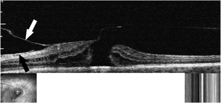
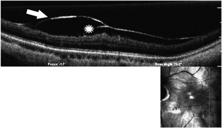
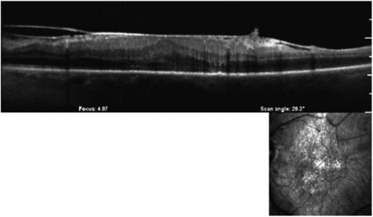
Methods: Histopathology was studied in 14 monkey eyes and a vitrectomy specimen of a patient with macular pucker. Optical coherence tomography/scanning laser ophthalmoscopy (OCT/SLO) was performed in 239 eyes: 45 MH, 45 MP, 51 dry age-related macular degeneration (AMD), 53 non-proliferative diabetic retinopathy (NPDR) and 45 controls.
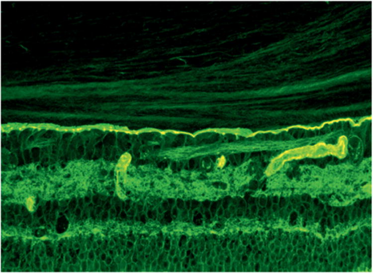
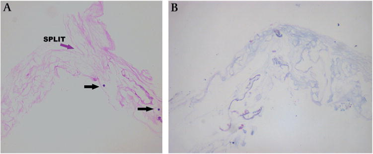
Results: Immunohistochemistry demonstrated lamellae in the posterior vitreous cortex of 12/14 (86%) monkey eyes. With OCT/SLO, vitreoschisis was detected in 24/45 (53%) MH and 19/45 (42%) MP eyes, but in only 7/53 (13%) NPDR, 3/51 (6%) AMD and 3/45 (7%) control eyes (p<0.001 for all comparisons). Rejoining of the inner and outer walls of the split posterior vitreous cortex was visible in 16/45 (36%) MH eyes and 15/45 (33%) MP eyes. Histopathology of the MP specimen confirmed a split with rejoining in the posterior vitreous cortex.
Conclusions: Vitreoschisis was detected in half of eyes with MH and MP, but much less frequently in controls, AMD and NPDR patients. These findings suggest that anomalous PVD with vitreoschisis may be pathogenic in MH and MP.
Conflict of interest statement
Figures
References
-
- Gass JD. Idiopathic senile macular hole. Its early stages and pathogenesis. Arch Ophthalmol. 1988;106:629–39. - PubMed
-
- Sebag J. Anomalous posterior vitreous detachment: a unifying concept in vitreoretinal disease. Graefes Arch Clin Exp Ophthalmol. 2004;242:690–8. - PubMed
-
- Roth AM, Foos RY. Surface wrinkling retinopathy in eyes enucleated at autopsy. Trans Am Acad Ophthalmol Otolaryngol. 1977;75:1047–58. - PubMed
-
- Sidd RJ, Fine SL, Owens SL, et al. Idiopathic preretinal gliosis. Am J Ophthalmol. 1982;94:44–8. - PubMed
-
- Vinores SA, Campochiaro PA, Conway BP. Ultrastructural and electron-immunocytochemical characterization of cells in epiretinal membranes. Invest Ophthalmol Vis Sci. 1990;31:14–28. - PubMed
Publication types
MeSH terms
Grants and funding
LinkOut - more resources
Full Text Sources