

Lysosomal iron mobilization and induction of the mitochondrial permeability transition in acetaminophen-induced toxicity to mouse hepatocytes
- PMID: 20584761
- PMCID: PMC2923283
- DOI: 10.1093/toxsci/kfq175
Lysosomal iron mobilization and induction of the mitochondrial permeability transition in acetaminophen-induced toxicity to mouse hepatocytes
Abstract
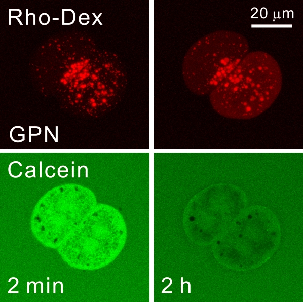
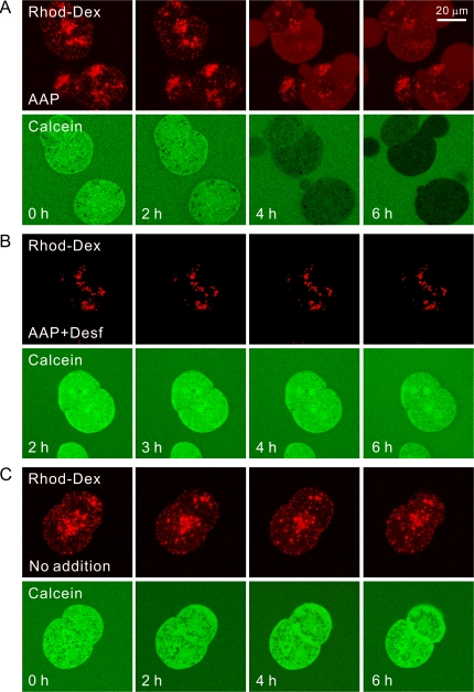
Acetaminophen induces the mitochondrial permeability transition (MPT) in hepatocytes. Reactive oxygen species (ROS) trigger the MPT and play an important role in AAP-induced hepatocellular injury. Because iron is a catalyst for ROS formation, our aim was to investigate the role of chelatable iron in MPT-dependent acetaminophen toxicity to mouse hepatocytes. Hepatocytes were isolated from fasted male C3Heb/FeJ mice. Necrotic cell killing was determined by propidium iodide fluorometry. Mitochondrial membrane potential was visualized by confocal microscopy of tetramethylrhodamine methylester. Chelatable ferrous ion was monitored by calcein quenching, and 70 kDa rhodamine-dextran was used to visualize lysosomes. Cell killing after acetaminophen (10mM) was delayed and decreased by more than half after 6 h by 1mM desferal or 1mM starch-desferal. In a cell-free system, ferrous but not ferric iron quenched calcein fluorescence, an effect reversed by dipyridyl, a membrane-permeable iron chelator. In hepatocytes loaded with calcein, intracellular calcein fluorescence decreased progressively beginning about 4 h after acetaminophen. Mitochondria then depolarized after about 6 h. Dipyridyl (20mM) dequenched calcein fluorescence. Desferal and starch-desferal conjugate prevented acetaminophen-induced calcein quenching and mitochondrial depolarization. As calcein fluorescence became quenched, lysosomes disappeared, consistent with release of iron from ruptured lysosomes. In conclusion, an increase of cytosolic chelatable ferrous iron occurs during acetaminophen hepatotoxicity, which triggers the MPT and cell killing. Disrupted lysosomes are the likely source of iron, and chelation of this iron decreases acetaminophen toxicity to hepatocytes.
Figures


Similar articles
-
Translocation of iron from lysosomes to mitochondria during acetaminophen-induced hepatocellular injury: Protection by starch-desferal and minocycline.Free Radic Biol Med. 2016 Aug;97:418-426. doi: 10.1016/j.freeradbiomed.2016.06.024. Epub 2016 Jun 23. Free Radic Biol Med. 2016. PMID: 27345134 Free PMC article.
-
Translocation of iron from lysosomes to mitochondria during ischemia predisposes to injury after reperfusion in rat hepatocytes.Free Radic Biol Med. 2013 Oct;63:243-53. doi: 10.1016/j.freeradbiomed.2013.05.004. Epub 2013 May 9. Free Radic Biol Med. 2013. PMID: 23665427 Free PMC article.
-
Translocation of iron from lysosomes into mitochondria is a key event during oxidative stress-induced hepatocellular injury.Hepatology. 2008 Nov;48(5):1644-54. doi: 10.1002/hep.22498. Hepatology. 2008. PMID: 18846543 Free PMC article.
-
The mitochondrial permeability transition in cell death: a common mechanism in necrosis, apoptosis and autophagy.Biochim Biophys Acta. 1998 Aug 10;1366(1-2):177-96. doi: 10.1016/s0005-2728(98)00112-1. Biochim Biophys Acta. 1998. PMID: 9714796 Review.
-
Mitochondrial dysfunction in the pathogenesis of necrotic and apoptotic cell death.J Bioenerg Biomembr. 1999 Aug;31(4):305-19. doi: 10.1023/a:1005419617371. J Bioenerg Biomembr. 1999. PMID: 10665521 Review.
Cited by
-
Acetaminophen-induced Liver Injury: from Animal Models to Humans.J Clin Transl Hepatol. 2014 Sep;2(3):153-61. doi: 10.14218/JCTH.2014.00014. Epub 2014 Sep 15. J Clin Transl Hepatol. 2014. PMID: 26355817 Free PMC article. Review.
-
Underlying mechanisms and treatment of acetaminophen‑induced liver injury (Review).Mol Med Rep. 2025 Apr;31(4):106. doi: 10.3892/mmr.2025.13471. Epub 2025 Feb 28. Mol Med Rep. 2025. PMID: 40017143 Free PMC article. Review.
-
Protective Activity of Total Polyphenols from Genista quadriflora Munby and Teucrium polium geyrii Maire in Acetaminophen-Induced Hepatotoxicity in Rats.Nutrients. 2016 Apr 1;8(4):193. doi: 10.3390/nu8040193. Nutrients. 2016. PMID: 27043622 Free PMC article.
-
The development and hepatotoxicity of acetaminophen: reviewing over a century of progress.Drug Metab Rev. 2020 Nov;52(4):472-500. doi: 10.1080/03602532.2020.1832112. Epub 2020 Oct 14. Drug Metab Rev. 2020. PMID: 33103516 Free PMC article. Review.
-
Recommendations for the use of the acetaminophen hepatotoxicity model for mechanistic studies and how to avoid common pitfalls.Acta Pharm Sin B. 2021 Dec;11(12):3740-3755. doi: 10.1016/j.apsb.2021.09.023. Epub 2021 Sep 30. Acta Pharm Sin B. 2021. PMID: 35024303 Free PMC article. Review.
References
-
- Badr MZ, Belinsky SA, Kauffman FC, Thurman RG. Mechanism of hepatotoxicity to periportal regions of the liver lobule due to allyl alcohol: role of oxygen and lipid peroxidation. J. Pharmacol. Exp. Ther. 1986;238:1138–1142. - PubMed
-
- Bajt ML, Knight TR, Lemasters JJ, Jaeschke H. Acetaminophen-induced oxidant stress and cell injury in cultured mouse hepatocytes: protection by N-acetyl cysteine. Toxicol. Sci. 2004;80:343–349. - PubMed
-
- Breuer W, Epsztejn S, Millgram P, Cabantchik IZ. Transport of iron and other transition metals into cells as revealed by a fluorescent probe. Am. J. Physiol. 1995;268(Pt 1):C1354–C1361. - PubMed
-
- Cohen SD, Khairallah EA. Selective protein arylation and acetaminophen-induced hepatotoxicity. Drug Metab. Rev. 1997;29:59–77. - PubMed
Publication types
MeSH terms
Substances
Grants and funding
LinkOut - more resources
Full Text Sources
Other Literature Sources
Medical
Research Materials
Miscellaneous
