

Hormonal suppression restores fertility in irradiated mice from both endogenous and donor-derived stem spermatogonia
- PMID: 20584762
- PMCID: PMC2923290
- DOI: 10.1093/toxsci/kfq191
Hormonal suppression restores fertility in irradiated mice from both endogenous and donor-derived stem spermatogonia
Abstract
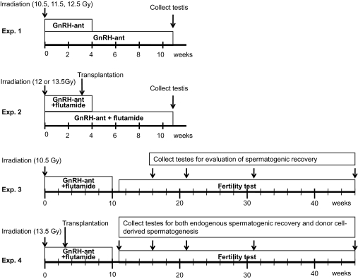
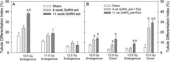
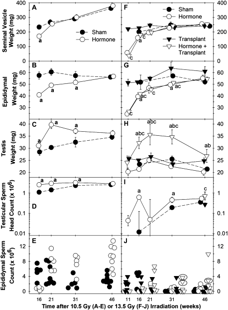
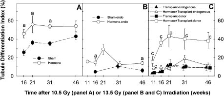
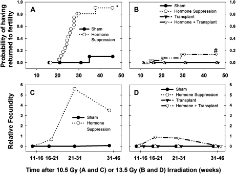
Irradiation interrupts spermatogenesis and causes prolonged sterility in male mammals. Hormonal suppression treatment with gonadotropin-releasing hormone (GnRH) analogues has restored spermatogenesis in irradiated rats, but similar attempts were unsuccessful in irradiated mice, monkeys, and humans. In this study, we tested a stronger hormonal suppression regimen (the GnRH antagonist, acyline, and plus flutamide) for efficacy both in restoring endogenous spermatogenesis and in enhancing colonization of transplanted stem spermatogonia in mouse testes irradiated with a total doses between 10.5 and 13.5 Gy. A 4-week hormonal suppression treatment, given immediately after irradiation, increased endogenous spermatogenic recovery 1.5-fold, and 11-week hormonal suppression produced twofold increases compared with sham-treated irradiated controls. Furthermore, 10-week hormonal suppression restored fertility from endogenous surviving spermatogonial stem cells in 90% of 10.5-Gy irradiated mice, whereas only 10% were fertile without hormonal suppression. Four- and 11-week hormonal suppression also enhanced spermatogenic development from transplanted stem spermatogonia in irradiated recipient mice, by 3.1- and 4.8-fold, respectively, compared with those not given hormonal treatment. Moreover, the 10-week hormonal suppression regimen, but not a sham treatment, restored fertility of some 13.5-Gy irradiated recipient mice from donor-derived spermatogonial stem cells. This is the first report of hormonal suppression inducing recovery of endogenous spermatogenesis and fertility in a mouse model treated with anticancer agents. The combination of spermatogonial transplantation with hormonal suppression should be investigated as a treatment to restore fertility in young men after cytotoxic cancer therapy.
Figures
Similar articles
-
Restoration of functional sperm production in irradiated pubertal rhesus monkeys by spermatogonial stem cell transplantation.Andrology. 2020 Sep;8(5):1428-1441. doi: 10.1111/andr.12807. Epub 2020 May 18. Andrology. 2020. PMID: 32351003 Free PMC article.
-
Hormone suppression with GnRH antagonist promotes spermatogenic recovery from transplanted spermatogonial stem cells in irradiated cynomolgus monkeys.Andrology. 2013 Nov;1(6):886-98. doi: 10.1111/j.2047-2927.2013.00126.x. Epub 2013 Sep 30. Andrology. 2013. PMID: 24124124 Free PMC article.
-
Effect of hormone modulations on donor-derived spermatogenesis or colonization after syngeneic and xenotransplantation in mice.Andrology. 2019 Mar;7(2):257-265. doi: 10.1111/andr.12571. Epub 2018 Nov 23. Andrology. 2019. PMID: 30471208 Free PMC article.
-
Hormonal approaches to preservation and restoration of male fertility after cancer treatment.J Natl Cancer Inst Monogr. 2005;(34):36-9. doi: 10.1093/jncimonographs/lgi002. J Natl Cancer Inst Monogr. 2005. PMID: 15784820 Review.
-
Suppression of testosterone stimulates recovery of spermatogenesis after cancer treatment.Int J Androl. 2003 Jun;26(3):141-6. doi: 10.1046/j.1365-2605.2003.00400.x. Int J Androl. 2003. PMID: 12755992 Review.
Cited by
-
Progress in germline stem cell transplantation in mammals and the potential usage.Reprod Biol Endocrinol. 2022 Mar 31;20(1):59. doi: 10.1186/s12958-022-00930-5. Reprod Biol Endocrinol. 2022. PMID: 35361229 Free PMC article. Review.
-
Restoration of functional sperm production in irradiated pubertal rhesus monkeys by spermatogonial stem cell transplantation.Andrology. 2020 Sep;8(5):1428-1441. doi: 10.1111/andr.12807. Epub 2020 May 18. Andrology. 2020. PMID: 32351003 Free PMC article.
-
Postpubertal spermatogonial stem cell transplantation restores functional sperm production in rhesus monkeys irradiated before and after puberty.Andrology. 2021 Sep;9(5):1603-1616. doi: 10.1111/andr.13033. Epub 2021 Jul 7. Andrology. 2021. PMID: 33960147 Free PMC article.
-
Single-Nucleus RNA-Seq Reveals Spermatogonial Stem Cell Developmental Pattern in Shaziling Pigs.Biomolecules. 2024 May 21;14(6):607. doi: 10.3390/biom14060607. Biomolecules. 2024. PMID: 38927011 Free PMC article.
-
Approaches and Technologies in Male Fertility Preservation.Int J Mol Sci. 2020 Jul 31;21(15):5471. doi: 10.3390/ijms21155471. Int J Mol Sci. 2020. PMID: 32751826 Free PMC article. Review.
References
-
- Boekelheide K, Schoenfeld H, Hall SJ, Weng CCY, Shetty G, Leith J, Harper J, Sigman M, Hess DL, Meistrich ML. Gonadotropin-releasing hormone antagonist (cetrorelix) therapy fails to protect non-human primates (macaca arctoides) from radiation-induced spermatogenic failure. J. Androl. 2005;26:222–234. - PubMed
-
- Crawford BA, Spaliviero JA, Simpson JM, Handelsman DJ. Testing the gonadal regression-cytoprotection hypothesis. Cancer Res. 1998;58:5105–5109. - PubMed
-
- Creemers LB, Meng X, Den Ouden K, Van Pelt AM, Izadyar F, Santoro M, Sariola H, De Rooij DG. Transplantation of germ cells from glial cell line-derived neurotrophic factor-overexpressing mice to host testes depleted of endogenous spermatogenesis by fractionated irradiation. Biol. Reprod. 2002;66:1579–1584. - PubMed
-
- da Cunha MF, Meistrich ML, Nader S. Absence of testicular protection by a gonadotropin releasing hormone analog against cyclophosphamide-induced testicular cytotoxicity in the mouse. Cancer Res. 1987;47:1093–1097. - PubMed
-
- de Rooij DG. Proliferation and differentiation of spermatogonial stem cells. Reproduction. 2001;121:347–354. - PubMed
Publication types
MeSH terms
Substances
Grants and funding
LinkOut - more resources
Full Text Sources
Medical
