. 2010 Aug;16(8):1516-29.
doi: 10.1261/rna.1833310. Epub 2010 Jun 28.

Identification and quantitative analyses of microRNAs located in the distal axons of sympathetic neurons

Affiliations

Identification and quantitative analyses of microRNAs located in the distal axons of sympathetic neurons

Orlangie Natera-Naranjo et al. RNA. 2010 Aug.

Abstract

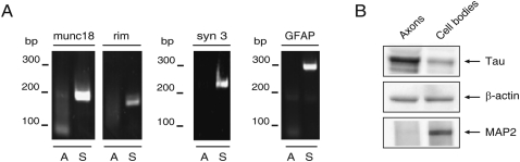
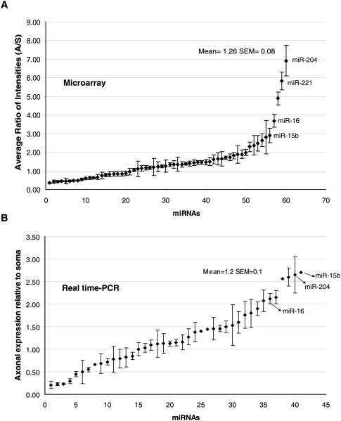
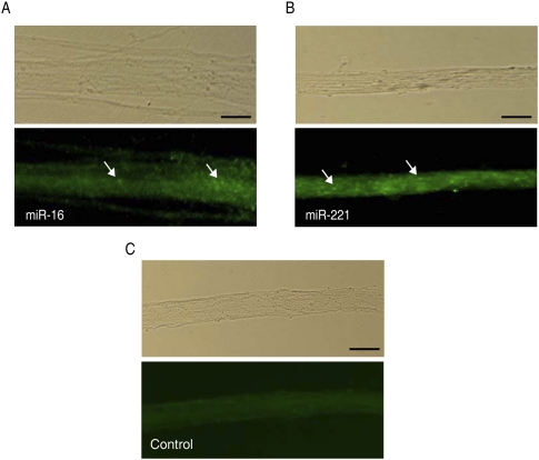
microRNAs (miRNAs) constitute a novel class of small, noncoding RNAs that act as negative post-transcriptional regulators of gene expression. Although the nervous system is a prominent site of miRNA expression, little is known about the spatial expression profiles of miRNAs in neurons. Here, we employed compartmentalized Campenot cell culture chambers to obtain a pure axonal RNA fraction of superior cervical ganglia (SCG) neurons, and determined the miRNA expression levels in these subcellular structural domains by microarray analysis and by real-time reverse-transcription polymerase chain reaction. The data revealed stable expression of a number of mature miRNAs that were enriched in the axons and presynaptic nerve terminals. Among the 130 miRNAs identified in the axon, miR-15b, miR-16, miR-204, and miR-221 were found to be highly abundant in distal axons as compared with the cell bodies of primary sympathetic neurons. Moreover, a number of miRNAs encoded by a common primary transcript (pri-miRNA) were differentially expressed in the distal axons, suggesting that there is a differential subcellular transport of miRNAs derived from the same coding region of the genome. Taken together, the data provide an important resource for future studies on the regulation of axonal protein synthesis and the role played by miRNAs in the maintenance of axonal structure and function as well as neuronal growth and development.


Figures

FIGURE 1.
FIGURE 1.
RNA prepared from the distal axons of SCG neurons is free of neuronal and glial contamination. (A) RT-PCR analysis was conducted on RNA obtained from the lateral and central compartments of the Campenot cell culture chambers using gene-specific primer sets for synaptic vesicle proteins, munc18, rim, synapsin 3 (syn 3), and glial fibrillary acidic protein (GFAP). PCR products were fractionated on 2.5% agarose gels and amplicons visualized by ethidium bromide staining. (A) Axon; (S) soma; (bp) base pairs. (B) Protein lysates obtained from the lateral and central compartments of the Campenot chambers were analyzed by Western blot using antibodies against Tau and MAP2. Levels of β-actin immunoreactivity served as an internal control.
FIGURE 2.
FIGURE 2.
A comparison of the abundance ratios (axon/soma) of miRNAs expressed in axons as judged by microarray analysis and qRT-PCR. (A) miRNA expression in the axons assessed by microarray. The average ratios of axonal miRNA intensities relative to miRNA intensity in the soma were calculated for 60 miRNAs. Error bars represent the standard error of mean (SEM) of six replicates. (B) miRNA expression in the axons assessed by real-time RT-PCR. Distribution plot shows the relative expression of axonal miRNAs compared with the soma as measured using miRNA-specific primers. Relative expression was calculated using the 2−ΔΔCT value after normalizing the CT values in the axons and soma to the corresponding levels of U6 snRNA. The distribution can be approximated as normal by means of the Shapiro-Wilk normality test (P = 0.286).
FIGURE 3.
FIGURE 3.
Visualization of miRNAs in distal axons by in situ hybridization (ISH). ISH was performed in primary SCG neurons at 7 d in vitro (DIV) using fluorescein isothiocyanate (FITC)-conjugated LNA probes for miR-16 (A), miR-221 (B), and a scrambled miR probe as a negative control (C). Arrows indicate the location of punctate ISH signals in distal axons for miR-16 and miR-221. Scale bars, 10 μm. Phase contrast images of the axon fibers are provided in the corresponding top panels.

Similar articles

Cited by

References

    1. Aldrich BT, Frakes EP, Kasuya J, Hammonda DL, Kitamoto T 2009. Changes in expression of sensory organ-specific microRNAs in rat dorsal root ganglia in association with mechanical hypersensitivity induced by spinal nerve ligation. Neuroscience 164: 711–723 - PMC - PubMed
    1. Ambros V, Lee RC 2004. Identification of microRNAs and other tiny noncoding RNAs by cDNA cloning. Methods Mol Biol 265: 131–158 - PubMed
    1. Aschrafi A, Schwechter AD, Mameza MG, Natera-Naranjo O, Gioio AE, Kaplan BB 2008. MicroRNA-338 regulates local cytochrome c oxidase IV mRNA levels and oxidative phosphorylation in the axons of sympathetic neurons. J Neurosci 28: 12581–12590 - PMC - PubMed
    1. Aschrafi A, Natera-Naranjo O, Gioio AE, Kaplan BB 2010. Regulation of axonal trafficking of cytochrome c oxidase IV mRNA. Mol Cell Neurosci 43: 422–430 - PMC - PubMed
    1. Bassell GJ, Warren ST 2008. Fragile X syndrome: Loss of local mRNA regulation alters synaptic development and function. Neuron 60: 201–214 - PMC - PubMed

Publication types

LinkOut - more resources