. 2010 Aug;16(8):1559-69.
doi: 10.1261/rna.2197210. Epub 2010 Jun 28.

The HCV IRES pseudoknot positions the initiation codon on the 40S ribosomal subunit

Affiliations

The HCV IRES pseudoknot positions the initiation codon on the 40S ribosomal subunit

Katherine E Berry et al. RNA. 2010 Aug.

Abstract

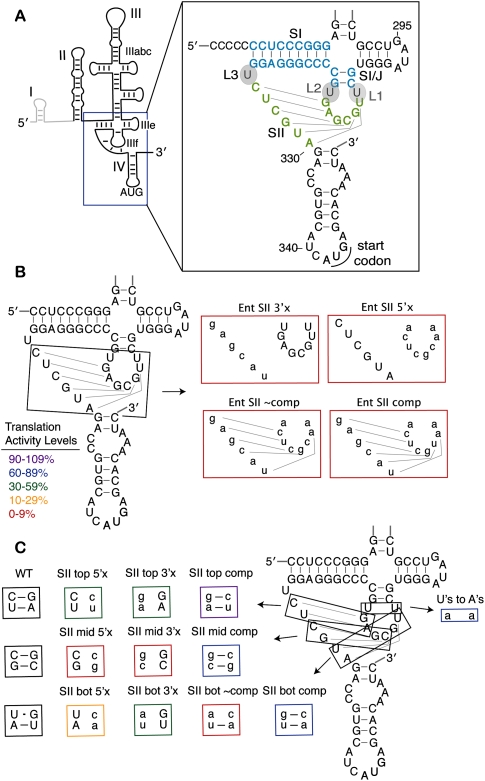
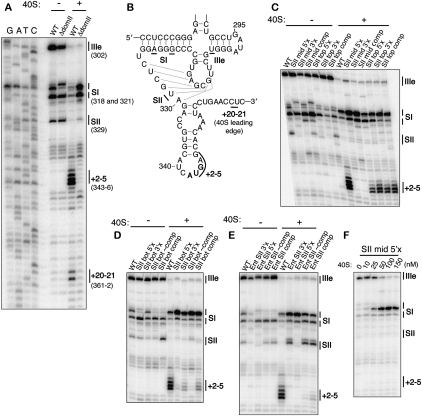
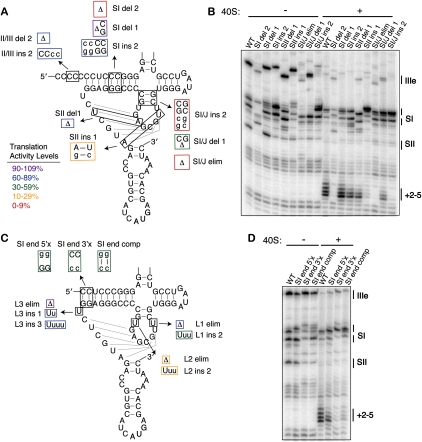
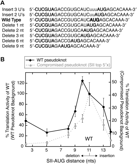
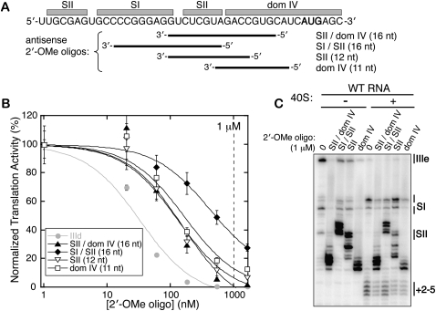
The hepatitis C virus (HCV) genomic RNA contains an internal ribosome entry site (IRES) in its 5' untranslated region, the structure of which is essential for viral protein translation. The IRES includes a predicted pseudoknot interaction near the AUG start codon, but the results of previous studies of its structure have been conflicting. Using mutational analysis coupled with activity and functional assays, we verified the importance of pseudoknot base pairings for IRES-mediated translation and, using 35 mutants, conducted a comprehensive study of the structural tolerance and functional contributions of the pseudoknot. Ribosomal toeprinting experiments show that the entirety of the pseudoknot element positions the initiation codon in the mRNA binding cleft of the 40S ribosomal subunit. Optimal spacing between the pseudoknot and the start site AUG resembles that between the Shine-Dalgarno sequence and the initiation codon in bacterial mRNAs. Finally, we validated the HCV IRES pseudoknot as a potential drug target using antisense 2'-OMe oligonucleotides.


Figures

FIGURE 1.
FIGURE 1.
Stem II base pairs form in the HCV IRES pseudoknot and contribute to IRES translation efficiency. (A) A line diagram of the predicted HCV IRES secondary structure with the major domains and the AUG start codon labeled. Dom I is shown in gray and is not included in the IRES-luciferase reporter constructs. The inset shows the sequence and predicted secondary structure of the pseudoknot with the stem and loop nomenclature indicated. SI is shown in light blue and SII is shown in light green. (B) Mutations of the entire (Ent) SII sequence, as previously reported in Kieft et al. (2001) and (C) mutations of SII made two base pairs at a time. Mutated nucleotides are shown in lowercase and the mutants are boxed in colors corresponding to their translation activity levels as defined in B. Disruptive mutations are named according to whether the 5′ or 3′ side of SII (in the primary sequence) was mutated to its complement.
FIGURE 2.
FIGURE 2.
Stem II base pairing contributes to AUG positioning by the IRES. (A) Establishment of primer extension and toeprinting stops for WT IRES in the absence and presence of 40S subunits and comparison to ΔdomII IRES. Primer extension stops were mapped based on dideoxy sequencing reactions. (B) Location of major primer extension stops mapped onto the pseudoknot predicted secondary structure. Primer extension inhibition and toeprinting of (C) the middle and top SII base pair mutants, (D) the bottom SII base pair mutants, and (E) the entire SII base pair mutants. (F) Primer extension reactions of SII mid-5′× IRES-FF luc mRNA (50 nM) in the presence of increasing concentrations of 40S ribosomal subunits. The positions of the major stops are indicated at the right of each gel and are defined in A and B.
FIGURE 3.
FIGURE 3.
Tolerance of the pseudoknot structure to stem and loop length. (A) Mutations of the pseudoknot stem lengths. Mutated nucleotides are shown in lowercase and the mutants are boxed in colors according to their translation activity, as defined in the legend. Deletions of nucleotides are shown as Δ. (B) Primer extension inhibition and toeprinting of stem length mutants. The positions of the major stops are indicated at the right of each gel and are defined in Figure 2, A and B. (C) Mutants of the pseudoknot loop lengths and base pairing at the terminus of SI, with the mutations and the activities represented as in A. (D) Primer extension inhibition and toeprinting of SI terminus base pair mutants, labeled as in B.
FIGURE 4.
FIGURE 4.
Correlation between toeprint strength and translation activity. Translation activity (as quantified in Table 1) plotted against toeprint stop intensity, quantified by densitometry, and normalized to the toeprint of the WT IRES. Plotted points reflect the mean value of activity across four in vitro translations from two independent transcriptions, and the mean value of the toeprint strength from two independent transcriptions. The error bars around the points extend 1 SD above and below the mean. Mutants are grouped into categories based on what structural features they disrupt, as indicated by the legend, but linear regression was conducted on the full set of 36 mutants together. The estimated slope with a standard error is 0.99 ± 0.11; the correlation is 0.84, which is significantly different from zero at the 0.0001 level.
FIGURE 5.
FIGURE 5.
Inhibition of IRES translation activity by varying the pseudoknot–AUG distance. (A) Sequences of mutations made to either lengthen or shorten the pseudoknot–AUG distance. Inserted nucleotides are shown in lowercase letters, and the start codon and SII are in bold. (B) Translation activity plotted against the pseudoknot–AUG distance with deletions plotted to the left of the WT AUG position and insertions to the right. Translation activities from the WT IRES background are plotted on the left vertical axis and those from a compromised pseudoknot (SII top 5′× mutant) on the right vertical axis, with 100% activity on the left axis scaled to 40% starting activity on the right. Plotted points represent the mean activities of four translation reactions from two independent transcriptions and the error bars extend 1 SD above and below the mean.
FIGURE 6.
FIGURE 6.
Inhibition of HCV IRES translation with pseudoknot-targeted 2′-OMe oligonucleotides. (A) The diagram shows to which regions of the IRES each of the four 2′-OMe oligonucleotides is complementary, with the start site AUG shown in bold. (B) Titrations of the 2′-OMe oligonucleotides in WT IRES-FF luciferase in vitro translation reactions, fit to sigmoidal inhibition curves. Plotted points represent the mean activities of four translation reactions from two independent transcriptions and the error bars extend 1 SD above and below the mean. The 1 μM concentration utilized in toeprinting experiments in C is indicated with a vertical dashed line. (C) Primer extension inhibition and toeprinting on the WT IRES-FF luc mRNA in the presence of 1 μM 2′-OMe oligonucleotides.

References

    1. Boehringer D, Thermann R, Ostareck-Lederer A, Lewis JD, Stark H 2005. Structure of the hepatitis C virus IRES bound to the human 80S ribosome: Remodeling of the HCV IRES. Structure 13: 1695–1706 - PubMed
    1. Brierley I, Pennell S, Gilbert RJ 2007. Viral RNA pseudoknots: Versatile motifs in gene expression and replication. Nat Rev Microbiol 5: 598–610 - PMC - PubMed
    1. Brown EA, Zhang H, Ping LH, Lemon SM 1992. Secondary structure of the 5′ nontranslated regions of hepatitis C virus and pestivirus genomic RNAs. Nucleic Acids Res 20: 5041–5045 - PMC - PubMed
    1. Deutsch M, Hadziyannis SJ 2008. Old and emerging therapies in chronic hepatitis C: An update. J Viral Hepat 15: 2–11 - PubMed
    1. Easton LE, Locker N, Lukavsky PJ 2009. Conserved functional domains and a novel tertiary interaction near the pseudoknot drive translational activity of hepatitis C virus and hepatitis C virus-like internal ribosome entry sites. Nucleic Acids Res 37: 5537–5549 - PMC - PubMed

Publication types