. 2010 Sep 10;285(37):28749-63.
doi: 10.1074/jbc.M110.148361. Epub 2010 Jun 28.

Bcl-2 and Bax interact via the BH1-3 groove-BH3 motif interface and a novel interface involving the BH4 motif

Affiliations

Bcl-2 and Bax interact via the BH1-3 groove-BH3 motif interface and a novel interface involving the BH4 motif

Jingzhen Ding et al. J Biol Chem. .

Abstract

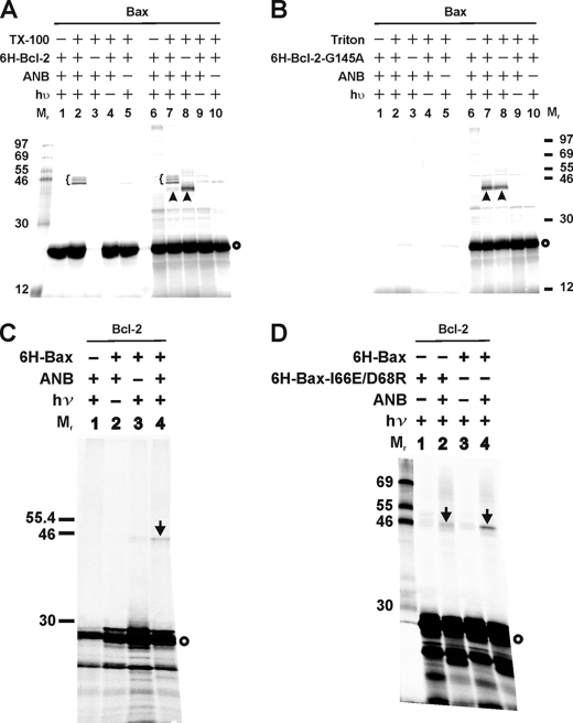
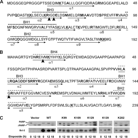
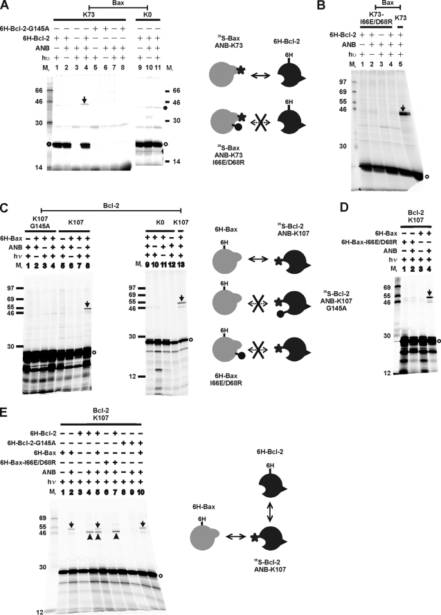
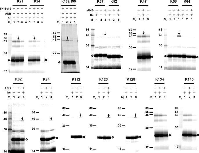
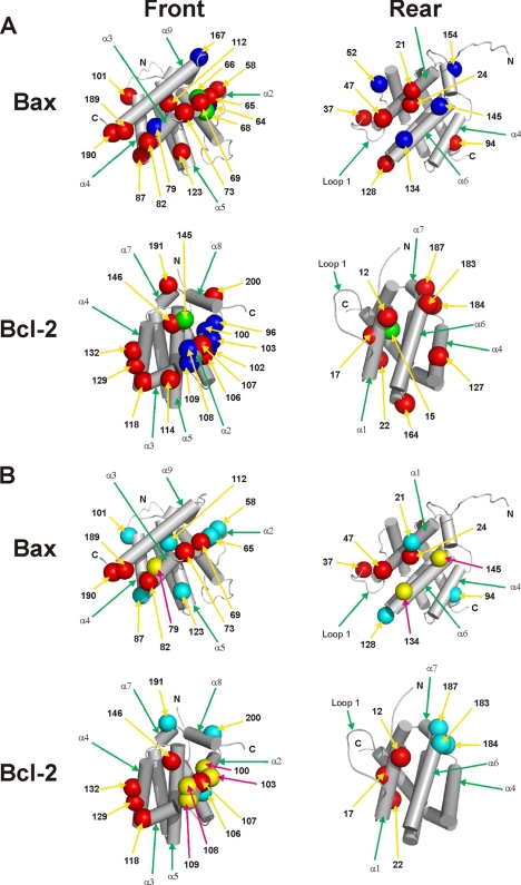
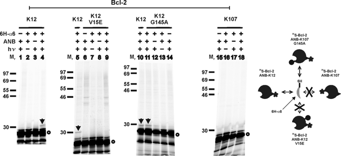
The interaction of Bcl-2 family proteins at the mitochondrial outer membrane controls membrane permeability and thereby the apoptotic program. The anti-apoptotic protein Bcl-2 binds to the pro-apoptotic protein Bax to prevent Bax homo-oligomerization required for membrane permeabilization. Here, we used site-specific photocross-linking to map the surfaces of Bax and Bcl-2 that interact in the hetero-complex formed in a Triton X-100 micelle as a membrane surrogate. Heterodimer-specific photoadducts were detected from multiple sites in Bax and Bcl-2. Many of the interaction sites are located in the Bcl-2 homology 3 (BH3) region of Bax and the BH1-3 groove of Bcl-2 that likely form the BH3-BH1-3 groove interface. However, other interaction sites form a second interface that includes helix 6 of Bax and the BH4 region of Bcl-2. Loss-of-function mutations in the BH3 region of Bax and the BH1 region of Bcl-2 disrupted the BH3-BH1-3 interface, as expected. Surprisingly the second interface was also disrupted by these mutations. Similarly, a loss-of-function mutation in the BH4 region of Bcl-2 that forms part of the second interface also disrupted both interfaces. As expected, both kinds of mutation abolished Bcl-2-mediated inhibition of Bax oligomerization in detergent micelles. Therefore, Bcl-2 binds Bax through two interdependent interfaces to inhibit the pro-apoptotic oligomerization of Bax.


Figures

FIGURE 1.
FIGURE 1.
Structures and interactions of Bax and Bcl-2. A, monomeric structures of Bax (left) and Bcl-2 (right) were drawn using PyMOL (DeLano Scientific) based on the coordinates 1F16 and 1G5M in the Protein Data Bank (4, 5). All α-helices and the loop between α1 and α2 are indicated. The front surface that includes the BH1–3 region is colored in medium gray. The rear surface formed by α1, the loop between α1 and α2, and α6 are colored in dark gray. The schematic below under each structure has the front and rear surfaces indicated and will be used in other figures to represent Bax and Bcl-2. B, model for Bcl-2 homo-complex formation that is proposed based on previous studies (36–39). Neighboring molecules in the homo-oligomers are shaded differently for clarity, although they have the same conformation and interaction. C, Bax and Bcl-2 can potentially bind through their front surfaces (left) or rear surfaces (right) to form heterodimers.
FIGURE 2.
FIGURE 2.
Photocross-linking detected Bax/Bcl-2 heterodimers. A, photocross-linking of in vitro synthesized ANB-Lys/[35S]Met-labeled Bax to His6-tagged Bcl-2ΔTM in the absence and presence of Triton X-100 (TX-100). Phosphorimaging data shown are from the total samples (lanes 6–10), and their corresponding Ni2+ resin-bound fractions (lanes 1–5). The heterodimer-specific photoadducts were detected in lanes 2–7 and are indicated by braces. A photoadduct formed by Bax and a protein in the reticulocyte lysate were detected in lanes 7 and 8 and indicated by arrowheads. In vitro synthesized [35S]Met-Bax monomer is indicated by a circle on the right of the phosphorimage. The molecular weight (Mr) of protein standards is indicated on the left. B, photocross-linking of the ANB-Lys/[35S]Met-labeled Bax to His6-tagged Bcl-2ΔTM-G145A (6H-Bcl-2-G145A). Both total (lanes 6–10) and the Ni2+ resin-bound (lanes 1–5) samples are shown. The symbols used to indicate the photoadduct of Bax and reticulocyte protein and the [35S]Met-Bax monomer are defined in A. C and D, photocross-linking of in vitro synthesized ANB-Lys/[35S]Met-labeled Bcl-2ΔTM to His6-tagged Bax or Bax-I66E/D68R. Data shown were from the Ni2+ resin-bound fractions of the samples. The heterodimer-specific photoadduct is indicated by an arrow, and the [35S]Met-Bcl-2ΔTM monomer by a circle.
FIGURE 3.
FIGURE 3.
Sequences of Bax and Bcl-2 mutants and anti-apoptotic activity of Bcl-2 mutants. Bax (A) and Bcl-2 (B) sequences are shown with BH motifs highlighted by dashed lines above and α-helices identified by arrows below. All nine lysines (underlined Ks) were changed to Arg to create Bax K0. Single Lys Bax mutants were created by replacing each of the residues highlighted in boldface with a Lys. Arrowheads indicate Ile66 and Asp68 of Bax that were changed to Glu and Arg, respectively, in the I66E/D68R mutant; and Val15 and Gly145 of Bcl-2 that were changed to Glu and Ala in the V15E and G145A mutants, respectively. The last 22 residues (in parentheses) were deleted in all Bcl-2ΔTM mutants. C, activity of wild type human Bcl-2 (WT) and the single Lys mutant proteins in Rat-1ERTAM cells. Cleavage of PARP to ΔPARP and expression of the Bcl-2 proteins in the etoposide-treated cells were followed by SDS-PAGE and immunoblotting of the cell extracts with PARP- (top panels) and Bcl-2 (bottom panels)-specific antibody, respectively. The Bcl-2 protein expressed stably in the cells is indicated above each panel. Cells were treated with etoposide for the time indicated below each panel prior to analysis. In the controls (vector and WT Bcl-2), the blot probed for human Bcl-2 was deliberately overexposed to show lack of background in the vector-transfected cells.
FIGURE 4.
FIGURE 4.
Site-specific photocross-linking detected Bax/Bcl-2 heterodimer. A, photocross-linking of in vitro synthesized [35S]Met-labeled Bax with a single ANB probe attached to K73 to His6-tagged Bcl-2ΔTM (lanes 1–4) or Bcl-2ΔTM-G145A (lanes 5–8). The control reactions with in vitro synthesized Bax K0 are shown in lanes 9–11. The photoadduct between the [35S]Met-Bax K73 and His6-Bcl-2ΔTM is indicated by an arrow. The adduct was absent when either His6-Bcl-2ΔTM-G145A (lane 8) or in vitro synthesized Bax K0 (lane 11) was used. The filled circle adjacent to lane 11 indicates a nonphotoadduct because it was also detected in the absence of UV irradiation (lane 10). The open circle indicates the [35S]Met-Bax K73 or K0 monomer. B, photocross-linking of the ANB/[35S]Met-labeled Bax K73-I66E/D68R to His6-tagged Bcl-2ΔTM. The control reaction with ANB/[35S]Met-labeled Bax K73 is shown in lane 5. C, photocross-linking of His6-tagged Bax to in vitro synthesized [35S]Met-labeled Bcl-2ΔTM with a single ANB probe attached to K107 (lanes 5–8). The heterodimer-specific photoadduct was detected in lane 8 and indicated by an arrow. The adduct was not detected when either Bcl-2ΔTM-G145A-K107 (lane 4) or Bcl-2ΔTM K0 (lane 9) was synthesized in vitro and used in the cross-linking reaction. The open circle indicates the [35S]Met-Bcl-2ΔTM monomer. D, photocross-linking of His6-tagged Bax-I66E/D68R with in vitro synthesized ANB/[35S]Met-labeled Bcl-2ΔTM K107. The control reaction with His6-Bax is shown in lane 4. E, competitive photocross-linking of His6-Bax and His6-Bcl-2ΔTM with the ANB/[35S]Met-labeled Bcl-2ΔTM K107. The photoadduct was formed between the ANB/[35S]Met-Bcl-2 K107 and His6-Bax or His6-Bcl-2ΔTM and indicated by an arrow or arrowhead, respectively. In all panels, the phosphorimaging data shown were from the Ni2+ resin-bound fractions of the samples. The schematics on the side of each panel illustrate the proteins (Bax, gray; Bcl-2, black) in the corresponding cross-linking reaction and their interaction (indicated by the double-headed arrow). The star indicates the ANB photocross-linker. The lollipop indicates a mutation that abolished the interaction (indicated by the double-headed arrow with cross).
FIGURE 5.
FIGURE 5.
Photocross-linking of His6-tagged Bcl-2ΔTM protein to [35S]Met-Bax mutant proteins, each with a single ANB-labeled lysine residue. The phosphorimaging data shown were from the Ni2+ resin-bound fractions of the samples. The position of the ANB-lysine in each [35S]Met-Bax mutant is indicated at the top of each image. The heterodimer-specific photoadduct and the [35S]Met-Bax mutant monomer are indicated by an arrow and circle, respectively.
FIGURE 6.
FIGURE 6.
Photocross-linking of His6-tagged Bax protein to [35S]Met-Bcl-2ΔTM mutant proteins, each with a single ANB-labeled lysine residue. The phosphorimaging data shown were from the Ni2+ resin-bound fractions of the samples. The position of the ANB-lysine in each [35S]Met-Bcl-2ΔTM mutant is indicated at the top of each image. The heterodimer-specific photoadduct and the [35S]Met-Bcl-2ΔTM mutant monomer are indicated by an arrow and circle, respectively.
FIGURE 7.
FIGURE 7.
Predicted interfaces of Bax-Bcl-2 hetero-complexes. A, based on the site-specific photocross-linking data shown above, the surfaces of Bax and Bcl-2 that may form the interface in the hetero-complex were modeled on the previously determined Bax and Bcl-2 structures (4, 5). The residues that, when replaced by ANB-Lys, generated a heterodimer-specific photoadduct and hence close to or within the interface are shown in red, and the residues that did not generate the photoadduct and hence likely far from the interface are shown in blue. The radii of these residues are increased to 6 Å to reflect the uncertainty of the photocross-linking-based mapping technique (36). The position of these residues in the sequence of corresponding protein is indicated by the number. The mutations, I66E/D68R in Bax and G145A and V15E in Bcl-2, are shown in green. B, surfaces of Bax and Bcl-2 that may form the interface in hetero-complex are overlaid with the surfaces that may form the interface in homo-complex according to our previous studies (36, 40). The red residues are involved in both homo- and hetero-interfaces, yellow residues in homo-interface only, and cyan residues in hetero-interface only. In all panels, the view on the left or right shows the front or rear surface, respectively. The residues assigned to each surface are those appeared in the corresponding surface in a space-filled structure that is oriented as the skeleton structure shown.
FIGURE 8.
FIGURE 8.
Photocross-linking of His6-tagged Bax peptide to [35S]Met/ANB-Lys-labeled Bcl-2ΔTM protein. The phosphorimaging data shown were from the Ni2+ resin-bound samples from cross-linking of [35S]Met-Bcl-2ΔTM protein with or without the indicated mutation via the ANB probe attached to K12 or K107 in the rear or front surface, respectively, with His6-tagged Bax helix 6 peptide (6H-α6) that is part of the rear surface of Bax. The position of the single lysine is indicated at the top. The photoadduct containing [35S]Met-Bcl-2ΔTM K12 and 6H-α6 is indicated by arrow. [35S]Met-Bcl-2ΔTM monomer is indicated by an open circle. The schematic on the right illustrates the cross-linking of the Bax peptide with corresponding Bcl-2 proteins that is dependent on the location of photoreactive probe and sensitive to one but not the other mutation.
FIGURE 9.
FIGURE 9.
Effect of mutation in the front or rear surface of Bax or Bcl-2ΔTM on heterodimer-specific cross-linking via photoreactive probe attached to the front or rear surface. Phosphorimaging data shown are from the Ni2+ resin-bound samples from cross-linking of [35S]Met-Bax with ANB attached to the indicated Lys to His6-Bcl-2ΔTM (A) or from cross-linking of [35S]Met-Bcl-2ΔTM with ANB attached to the indicated Lys to His6-Bax (B and C). In these panels, the arrow indicates the corresponding photoadduct, and the open circle indicates the monomer of 35S-labeled Bax or Bcl-2ΔTM protein. Mutant Bax and Bcl-2 were used if indicated. The arrowhead-indicated band in lane 14 of C is not a photoadduct with His6-Bax because it was also detected in the absence of His6-Bax (lane 11). D, schematic illustration of the corresponding proteins used in the cross-linking reactions here and in Fig. 4 with the locations of photoreactive probe and mutation, and the effect of mutation is indicated. The information is also summarized in Tables 3 and 4.
FIGURE 10.
FIGURE 10.
Effect of the interface mutations on Bcl-2 inhibition of Bax oligomerization in Triton X-100. Oligomerization of Triton X-100-treated His6-Bax or the I66E/D68R mutant in the absence or presence of His6-tagged Bcl-2ΔTM or the V15E mutant examined by gel filtration chromatography. The chromatographic fractions 21–32 were analyzed by SDS-PAGE and immunoblotting with a Bax- or Bcl-2-specific antibody to detect His6-tagged Bax (left panels) or Bcl-2 (right panels). The molar ratio of Bax versus Bcl-2 in the Bcl-2 containing sample is 1:6. The elution positions for protein standards are indicated at the top with the molecular weight. The predicted molecular weight for Bax or Bcl-2ΔTM monomer in a CHAPS micelle is 27 or 31, respectively. Thus, the WT and mutant Bax proteins are mainly in dimeric and oligomeric forms under all of the conditions, whereas the WT and mutant Bcl-2ΔTM proteins are in monomeric and dimeric forms.

Similar articles

Cited by

References

    1. Leber B., Lin J., Andrews D. W. (2007) Apoptosis 12, 897–911 - PMC - PubMed
    1. Youle R. J., Strasser A. (2008) Nat. Rev. Mol. Cell Biol. 9, 47–59 - PubMed
    1. Kvansakul M., Yang H., Fairlie W. D., Czabotar P. E., Fischer S. F., Perugini M. A., Huang D. C., Colman P. M. (2008) Cell Death Differ. 15, 1564–1571 - PubMed
    1. Petros A. M., Medek A., Nettesheim D. G., Kim D. H., Yoon H. S., Swift K., Matayoshi E. D., Oltersdorf T., Fesik S. W. (2001) Proc. Natl. Acad. Sci. U.S.A. 98, 3012–3017 - PMC - PubMed
    1. Suzuki M., Youle R. J., Tjandra N. (2000) Cell 103, 645–654 - PubMed

Publication types

MeSH terms

Substances