. 2010 Sep 3;285(36):27869-78.
doi: 10.1074/jbc.M110.133066. Epub 2010 Jun 28.

Activation of Na+/H+ exchanger NHE3 by angiotensin II is mediated by inositol 1,4,5-triphosphate (IP3) receptor-binding protein released with IP3 (IRBIT) and Ca2+/calmodulin-dependent protein kinase II

Affiliations

Activation of Na+/H+ exchanger NHE3 by angiotensin II is mediated by inositol 1,4,5-triphosphate (IP3) receptor-binding protein released with IP3 (IRBIT) and Ca2+/calmodulin-dependent protein kinase II

Peijian He et al. J Biol Chem. .

Abstract

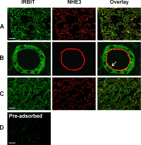
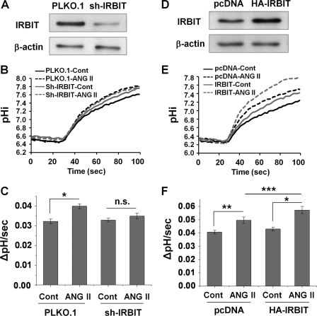
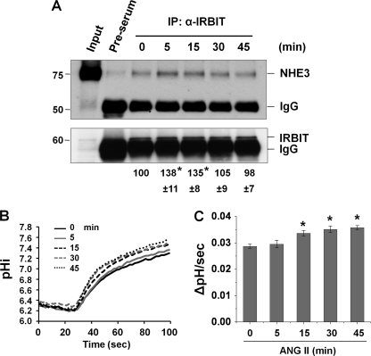
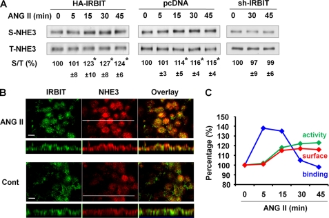
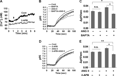
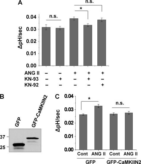
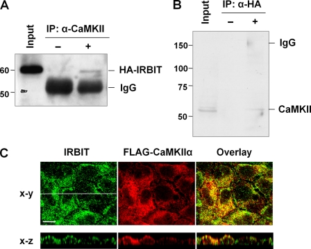
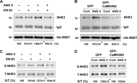
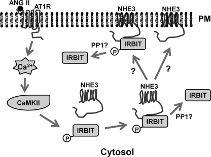
Angiotensin II (ANG II) stimulates renal tubular reabsorption of NaCl by targeting Na(+)/H(+) exchanger NHE3. We have shown previously that inositol 1,4,5-triphosphate receptor-binding protein released with inositol 1,4,5-triphosphate (IRBIT) plays a critical role in stimulation of NHE3 in response to elevated intracellular Ca(2+) concentration ([Ca(2+)](i)). In this study, we investigated the role of IRBIT in mediating NHE3 activation by ANG II. IRBIT is abundantly expressed in the proximal tubules where NHE3 is located. ANG II at physiological concentrations stimulates NHE3 transport activity in a model proximal tubule cell line. ANG II-induced activation of NHE3 was abrogated by knockdown of IRBIT, whereas overexpression of IRBIT enhanced the effect of ANG II on NHE3. ANG II transiently increased binding of IRBIT to NHE3 at 5 min but became dissociated by 45 min. In comparison, it took at least 15 min of ANG II treatment for an increase in NHE3 activity and NHE3 surface expression. The stimulation of NHE3 by ANG II was dependent on changes in [Ca(2+)](i) and Ca(2+)/calmodulin-dependent protein kinases II. Inhibition of CaMKII completely blocked the ANG II-induced binding of IRBIT to NHE3 and the increase in NHE3 surface abundance. Several serine residues of IRBIT are thought to be important for IRBIT binding. Mutations of Ser-68, Ser-71, and Ser-74 of IRBIT decreased binding of IRBIT to NHE3 and its effect on NHE3 activity. In conclusion, our current findings demonstrate that IRBIT is critically involved in mediating activation of NHE3 by ANG II via a Ca(2+)/calmodulin-dependent protein kinases II-dependent pathway.


Figures

FIGURE 1.
FIGURE 1.
Expression of IRBIT and NHE3 in the rat kidney. Cellular distribution of IRBIT and NHE3 in the rat kidney was determined by indirect fluorescence microscopic analysis using an affinity-purified polyclonal anti-IRBIT antibody and a monoclonal anti-NHE3 antibody, respectively. A, IRBIT (green) and NHE3 (red) expression in the cortex is shown. B, enlarged view of the proximal tubule. Arrow indicates co-localization of IRBIT with NHE3 at the luminal membrane. C, IRBIT (green) and NHE3 (red) expression in outer medulla is shown. D, immunofluorescence labeling of a cortical section was performed using an anti-IRBIT antibody that was preadsorbed with the recombinant IRBIT peptide antigen (16). Scale bar, 20 μm.
FIGURE 2.
FIGURE 2.
IRBIT is necessary for ANG II-mediated activation of NHE3 in OKP cells. A, OKP cells were infected with lentivirus carrying shRNA targeting IRBIT (sh-IRBIT) or the lentivirus PLKO.1 as a control. Western blot shows the expression level of IRBIT. β-Actin was used as a loading control. B, representative traces of Na+-dependent pHi recovery in OKP/PLKO.1 and OKP/sh-IRBIT cells treated with or without 1 nm ANG II for 45 min. Cont, control. C, NHE3 activity in response to 1 nm ANG II for 45 min was determined in OKP/PLKO.1 and OKP/sh-IRBIT cells. The rates of pHi recovery, ΔpH/s, at pHi 6.8 are shown. n = 10 or more. *, p < 0.001 compared with the control. n.s., not significant. D, expression level of IRBIT in OKP cells transfected with pcDNA/HA-IRBIT (HA-IRBIT) or a control vector (pcDNA) is shown. E, representative traces of Na+-dependent pHi recovery in OKP/pcDNA and OKP/HA-IRBIT treated with or without 1 nm ANG II for 45 min. F, NHE3 activities in response to 1 nm ANG II for 45 min were determined in OKP/pcDNA and OKP/HA-IRBIT cells. The rates of pHi recovery, ΔpH/s, at pHi 6.6 are shown. n = 10 or more. *, p < 0.001; **, p < 0.01; ***, p < 0.05.
FIGURE 3.
FIGURE 3.
NHE3-IRBIT interaction is stimulated by ANG II. A, OKP cells were treated with 1 nm ANG II for 0–45 min, and the cell lystes were incubated with an anti-IRBIT antibody or a pre-serum. Upper panel, the amount of co-immunoprecipitated NHE3 was examined by Western blot with an anti-NHE3 antibody. Lower panel, total amount of immunoprecipitated (IP) IRBIT determined using an anti-IRBIT antibody is shown. The amount of co-immunoprecipitated NHE3 was normalized to the total amount of IRBIT, and the relative changes are shown below the immunoblots. Data are represented as means ± S.E. of three independent experiments. *, p < 0.01 compared with 0 min. B, representative traces of Na+-dependent pHi recovery in OKP cells treated with ANG II for 0–45 min. C, NHE3 activity in response to ANG II was determined in OKP cells for up to 45 min. The rates of pHi recovery, ΔpH/s, at pH 6.6 are shown. n = 8 or more. *, p < 0.001 compared with 0 min.
FIGURE 4.
FIGURE 4.
ANG II increases NHE3 protein abundance in the apical membrane. A, OKP/pcDNA, OKP/HA-IRBIT, or OKP/sh-IRBIT cells were treated with ANG II for 0–45 min, and the amount of NHE3 on the apical membrane was determined by surface biotinylation as detailed under “Experimental Procedures.” Aliquots of surface and total protein were resolved by SDS-PAGE, and the amounts of surface (S) and total (T) NHE3 proteins were determined by Western blot using an anti-NHE3 antibody. The amount of surface NHE3 was normalized to total NHE3, and the relative changes are shown as the mean ± S.E. of at least three independent experiments (n = 3 for OKP/HA-IRBIT and OKP/sh-IRBT, n = 6 for OKP/pcDNA). *, p < 0.01 compared with 0 min. B, OKP/HA-IRBIT cells were treated with or without 1 nm ANG II for 45 min, and the expression of NHE3 (red; anti-NHE3) and IRBIT (green; anti-IRBIT) was determined by indirect immunofluorescence confocal microscopy. Confocal focal planes (x-y) and cross-sections (x-z) are shown. Bar, 10 μm. Representative images from three independent experiments are shown. C, summary of the changes in NHE3-IRBIT binding, NHE3 activity, and NHE3 surface expression in OKP cells is shown.
FIGURE 5.
FIGURE 5.
Activation of NHE3 by ANG II is Ca2+-dependent. A, OKP cells were pretreated with or without 50 μm 2-APB during Fura-2 loading. Changes in [Ca2+]i in response to 1 nm ANG II were determined by ratiometric measurements. Arrow indicates where ANG II was applied. Representative images are shown. n = 16 or more. OKP cells were pretreated with 20 μm BAPTA (B and C) or 50 μm 2-APB (D and E) for 20 min, and NHE3 activities were determined after 45 min of treatment with or without 1 nm ANG II. B and D, representative traces of Na+-dependent pHi recovery. C and E, NHE3 activity is presented as ΔpH/s, and the pHi recovery rates at pHi 6.8 are shown. n = 8 or more. *, p < 0.001; **, p < 0.01; n.s., not significant; Cont, control.
FIGURE 6.
FIGURE 6.
Activation of NHE3 by ANG II is CaMKII-dependent. A, OKP cells were pretreated with KN-93 or KN-92 for 20 min, and NHE3 activity was measured after 45 min of treatment with or without 1 nm ANG II. The rates of pHi recovery at pH 6.6 are shown. *, p < 0.01; n.s., not significant. B, OKP cells were transfected with pEGFP/CaMKIIN2 or pEGFP as a control (Cont). Western blot shows expression of GFP or GFP-CaMKIIN2. C, NHE3 activity in response to ANG II was determined in OKP/pEGFP and OKP/CaMKIIN2. The rates of pHi recovery, ΔpH/s, at pH 6.8 are shown. n = 8 or more. *, p < 0.001; n.s., not significant.
FIGURE 7.
FIGURE 7.
IRBIT interacts with CaMKII in OKP cells. A, cell lysates from OKP/HA-IRBIT cells were incubated with an anti-CaMKII antibody (+) or a rabbit serum (−), and co-immunoprecipitated (IP) IRBIT was detected by immunoblotting using an anti-HA antibody. B, cell lysates from OKP/HA-IRBIT cells were incubated with or without an anti-HA antibody, and the presence of co-immunoprecipitated CaMKII was determined. C, OKP cells were ectopically expressed with FLAG-CaMKIIα, and subcellular localization of IRBIT and CaMKIIα were determined by indirect immunofluorescence with anti-IRBIT and anti-FLAG antibodies, respectively. Representative images of focal (x-y) and cross-sectional (x-z) views from three independent experiments are shown. Bar, 10 μm.
FIGURE 8.
FIGURE 8.
ANG II-mediated IRBIT-NHE3 interaction and trafficking of NHE3 are CaMKII-dependent. Inhibition of CaMKII was achieved by KN-93 (A and C) or CaMKIIN2 (B and D). A and B, interaction between NHE3 and IRBIT was determined by treating cells with ANG II for 5 min. Co-immunoprecipitation of NHE3 with HA-IRBIT was determined by Western blotting. The amount of co-immunoprecipitated NHE3 was normalized to the total amount of IRBIT, and data are represented as means ± S.E. of three independent experiments. *, p < 0.01. C and D, NHE3 protein expression in the apical membrane was determined by surface biotinylation in cells treated with ANG II for 45 min. Aliquots of surface and total protein were resolved by SDS-PAGE, and the amount of surface (S) NHE3 and total (T) NHE3 proteins were determined by Western blot using an anti-NHE3 antibody. The amounts of surface NHE3 were normalized to total NHE3, and the relative changes are shown below the Western blots as means ± S.E. of three independent experiments. *, p < 0.01; Cont, control.
FIGURE 9.
FIGURE 9.
NHE3-IRBIT interaction and activation of NHE3 by ANG II are dependent on phosphorylation of IRBIT. OKP cells were ectopically expressed with HA-tagged wild-type IRBIT (WT), S68A, or S68A/S71A/S74A IRBIT mutants. A, cell lysates were incubated with an anti-HA antibody, and the co-immunoprecipitated NHE3 is shown in the upper panel. Immunoprecipitated (IP) HA-IRBIT and NHE3 and HA-IRBIT in the lysate are shown in the lower panels. The relative amounts of co-immunoprecipitated NHE3 are indicated below the immunoblots. Data are represented as mean ± S.E. of three independent experiments. *, p < 0.001. B, NHE3 activity was determined in OKP cells expressing WT or mutant HA-IRBIT after exposure to ANG II or carrier for 45 min. n = 8 or more. Percent increases in the rate of pHi recovery at pHi 6.8 are shown above the vertical bars. *, p < 0.01 comparing OKP/pcDNA + ANG II and OKP/WT + ANG II.
FIGURE 10.
FIGURE 10.
Putative model for ANG II-mediated activation of NHE3. Activation of angiotensin receptor (AT1R) induces mobilization of intracellular Ca2+ and subsequent activation of CaMKII. CaMKII phosphorylates IRBIT enhancing its interaction between NHE3 and trafficking of NHE3 to the apical membrane. PP1, protein phosphatase 1.

Similar articles

Cited by

References

    1. Biemesderfer D., Pizzonia J., Abu-Alfa A., Exner M., Reilly R., Igarashi P., Aronson P. S. (1993) Am. J. Physiol. 265, F736–F742 - PubMed
    1. Aronson P. S. (1996) Kidney Int. 49, 1665–1670 - PubMed
    1. Schultheis P. J., Clarke L. L., Meneton P., Miller M. L., Soleimani M., Gawenis L. R., Riddle T. M., Duffy J. J., Doetschman T., Wang T., Giebisch G., Aronson P. S., Lorenz J. N., Shull G. E. (1998) Nat. Genet. 19, 282–285 - PubMed
    1. Brown G. P., Douglas J. G. (1982) Endocrinology 111, 1830–1836 - PubMed
    1. Liu F. Y., Cogan M. G. (1988) J. Clin. Invest. 82, 601–607 - PMC - PubMed

Publication types

MeSH terms