. 2010 Jun 28;189(7):1107-15.
doi: 10.1083/jcb.201001149.

Vinculin potentiates E-cadherin mechanosensing and is recruited to actin-anchored sites within adherens junctions in a myosin II-dependent manner

Affiliations

Vinculin potentiates E-cadherin mechanosensing and is recruited to actin-anchored sites within adherens junctions in a myosin II-dependent manner

Quint le Duc et al. J Cell Biol. .

Erratum in

  • J Cell Biol. 2010 Nov 15;191(4):891

Abstract

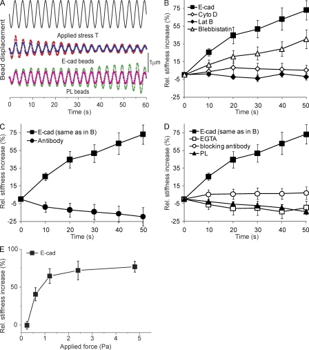
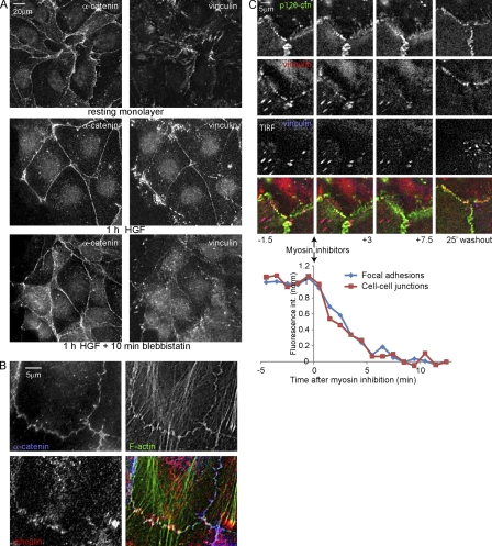
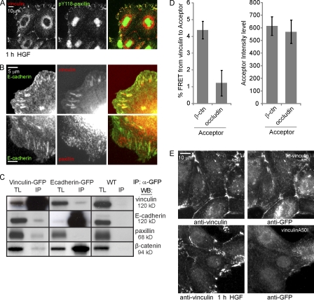
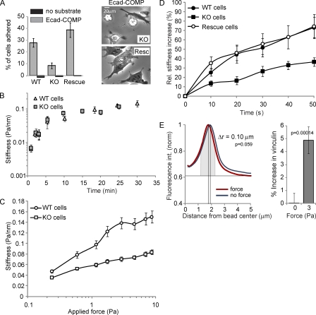
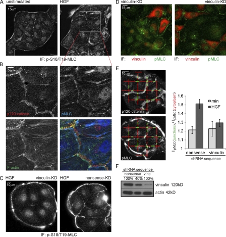
Cell surface receptors integrate chemical and mechanical cues to regulate a wide range of biological processes. Integrin complexes are the mechanotransducers between the extracellular matrix and the actomyosin cytoskeleton. By analogy, cadherin complexes may function as mechanosensors at cell-cell junctions, but this capacity of cadherins has not been directly demonstrated. Furthermore, the molecular composition of the link between E-cadherin and actin, which is needed to sustain such a function, is unresolved. In this study, we describe nanomechanical measurements demonstrating that E-cadherin complexes are functional mechanosensors that transmit force between F-actin and E-cadherin. Imaging experiments reveal that intercellular forces coincide with vinculin accumulation at actin-anchored cadherin adhesions, and nanomechanical measurements show that vinculin potentiates the E-cadherin mechanosensory response. These investigations directly demonstrate the mechanosensory capacity of the E-cadherin complex and identify a novel function for vinculin at cell-cell junctions. These findings have implications for barrier function, morphogenesis, cell migration, and invasion and may extend to all soft tissues in which classical cadherins regulate cell-cell adhesion.


Figures

Figure 1.
Figure 1.
E-cadherin is a mechanosensor. (A) Continuous driving field modulation at 0.3 Hz for 60 s (2.4 Pa stress) and representative time course of the displacement of two E-cadherin– and poly-l-lysine (PL)–coated beads. (B) The force-induced stiffening of Fc–E-cadherin–coated beads relative to unperturbed bead–cell contacts in the absence (E-cad) or presence of latrunculin B (Lat B), cytochalasin D (Cyto D), or blebbistatin. (C) Fc–E-cadherin–coated beads versus beads coated with monoclonal E-cadherin antibody. (D) Fc–E-cadherin–coated beads in the absence or presence of 3 mM EGTA added just before MTC or blocking anti–E-cadherin antibody versus beads coated with poly-l-lysine. (E) The percent change in E-cadherin junction stiffness relative to unperturbed cells as a function of applied shear stress is shown. After 20 min of bead–cell contact, the beads were subjected to a modulated 0.3-Hz magnetic field for 60 s. The elastic shear modulus was determined at 50 s as a function of the amplitude of the applied shear stress. (B–E) Each data point represents >300 beads. Error bars represent SD.
Figure 2.
Figure 2.
Vinculin is recruited to active cell–cell junctions in a myosin-dependent manner. (A) IF after CSK buffer extraction shows HGF-induced and myosin II–dependent α-catenin and vinculin distribution. (B) Magnified view of vinculin in cell–cell junctions of HGF-stimulated cells. (C) Cells expressing EGFP-vinculin (stably) and mCherry–p120-catenin (p120-ctn; transiently) analyzed by widefield and TIRF microscopy 1 h after HGF treatment. 3 µM ML-7 and 10 µM Y27632 were added 15 min after the start of imaging and washed out after another 10 min. EGFP-vinculin fluorescence intensity was measured in FAs (15 ROIs, each containing two to four FAs from seven time lapses) and cell–cell contacts (eight ROIs from seven time lapses).
Figure 3.
Figure 3.
Vinculin closely interacts with the E-cadherin complex. (A) Recruitment of vinculin but not pY118-paxillin to cell–cell junctions in HGF-stimulated cells shown by IF. (B) Colocalization of vinculin but not paxillin with GFP–E-cadherin in E-cadherin–COMP adhesions revealed by IF. (C) Western blot (WB) analyses of total lysates (TL) and IP of endogenous E-cadherin, vinculin, paxillin, and β-catenin co-IP with GFP-tagged vinculin or E-cadherin from cell lysates after cross-linking. Black lines indicate that intervening lanes have been spliced out. (D, left) FRET from immunolabeled GFP-vinculin (Alexa Fluor 488) to immunolabeled β-catenin (β-ctn) or occludin (rhodamine). Error bars represent SEM (n = 18). (right) Mean acceptor fluorescence intensity in the ROIs used for calculating FRET. (E) MDCK cells transfected with GFP-vinculin or GFP–vinculin A50I were washed with CSK buffer, fixed, and immunostained for vinculin and GFP simultaneously.
Figure 4.
Figure 4.
Vinculin modulates E-cadherin mechanosensing. (A, left) The relative number of wt, ko, and vinculin-reconstituted F9 cells that adhered to E-cadherin–COMP-coated wells after 45 min is shown. (right) Phase-contrast images of ko and reconstituted cells on E-cadherin–COMP. (B) The force-independent stiffening of E-cadherin junctions as a function of the bead–cell contact time. After increasing periods of bead–cell contact, the oscillating field (0.3 Hz at 10 Gauss) was switched on for 10 s to quantify the elastic shear modulus (Pa/nm). (C) Junctional stiffness in wt versus ko cells in response to increasing applied shear stress. After 20 min of bead–cell contact, the field strength was increased stepwise in 10-s intervals with no pause between successive changes in the magnetic field. Each data point represents the mean. (D) The force-induced stiffening of Fc–E-cadherin–coated beads bound for 20 min to wt, ko, and vinculin-reconstituted F9 cells was measured using a modulated 0.3-Hz field (20 Gauss) for 50 s. (E, left) The mean intensity profile of vinculin IF plotted against the distance from the bead center for ∼80 unforced and forced beads. (right) Total vinculin intensity above baseline at unforced and forced beads measured in a 1-µm-wide area around the maximum of fluorescence intensity (gray). Error bars represent SD (A, triplicates; B–D, n > 300 [approximately one bead/cell]; E, n = ∼80).
Figure 5.
Figure 5.
Vinculin knockdown prevents HGF-induced pMLC accumulation near cell–cell junctions. (A) HGF (2 h) induced increase in pMLC near cell–cell junctions revealed by IF. (B) A magnified view of A, showing that pMLC accumulates at F-actin structures that connect to p120-labeled cell–cell junctions. (C) IF shows a reduction of pMLC recruited to cell–cell junctions in vinculin knockdown (KD) cells after 2 h of HGF. (D) Overlay of the vinculin channel (smoothed with a Gaussian; r = 40) and pMLC channel of a dual-color IF staining of vinculin knockdown cells after 2 h of HGF. (E, left) Representative image automatically generated by custom software, displaying the line fragments used to measure pMLC levels near cell–cell junctions (green) and in the cytoplasm (red). (right) The relative intensity of pMLC near cell–cell junctions (a value of 1 means equal levels) shows an increase after 2 h of HGF that is largely abolished by vinculin knockdown. Error bars represent SEM. 25 images were analyzed for each condition.

Comment in

Similar articles

Cited by

References

    1. Abe K., Takeichi M. 2008. EPLIN mediates linkage of the cadherin catenin complex to F-actin and stabilizes the circumferential actin belt. Proc. Natl. Acad. Sci. USA. 105:13–19 10.1073/pnas.0710504105 - DOI - PMC - PubMed
    1. Avizienyte E., Fincham V.J., Brunton V.G., Frame M.C. 2004. Src SH3/2 domain-mediated peripheral accumulation of Src and phospho-myosin is linked to deregulation of E-cadherin and the epithelial-mesenchymal transition. Mol. Biol. Cell. 15:2794–2803 10.1091/mbc.E03-12-0879 - DOI - PMC - PubMed
    1. Ayollo D.V., Zhitnyak I.Y., Vasiliev J.M., Gloushankova N.A. 2009. Rearrangements of the actin cytoskeleton and E-cadherin-based adherens junctions caused by neoplasic transformation change cell-cell interactions. PLoS One. 4:e8027 10.1371/journal.pone.0008027 - DOI - PMC - PubMed
    1. Bakolitsa C., Cohen D.M., Bankston L.A., Bobkov A.A., Cadwell G.W., Jennings L., Critchley D.R., Craig S.W., Liddington R.C. 2004. Structural basis for vinculin activation at sites of cell adhesion. Nature. 430:583–586 10.1038/nature02610 - DOI - PubMed
    1. Bershadsky A.D., Balaban N.Q., Geiger B. 2003. Adhesion-dependent cell mechanosensitivity. Annu. Rev. Cell Dev. Biol. 19:677–695 10.1146/annurev.cellbio.19.111301.153011 - DOI - PubMed

Publication types