. 2010 Sep;30(17):4234-44.
doi: 10.1128/MCB.01396-09. Epub 2010 Jun 28.

Islet beta-cell-specific MafA transcription requires the 5'-flanking conserved region 3 control domain

Affiliations

Islet beta-cell-specific MafA transcription requires the 5'-flanking conserved region 3 control domain

Jeffrey C Raum et al. Mol Cell Biol. 2010 Sep.

Abstract

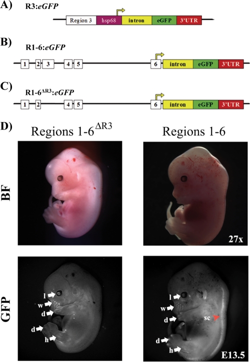
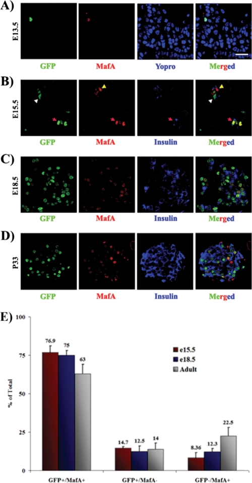
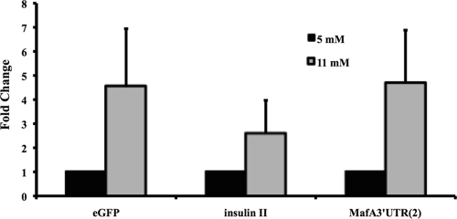
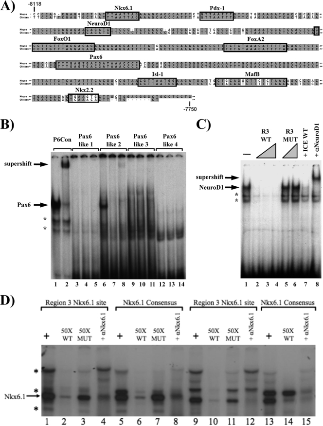
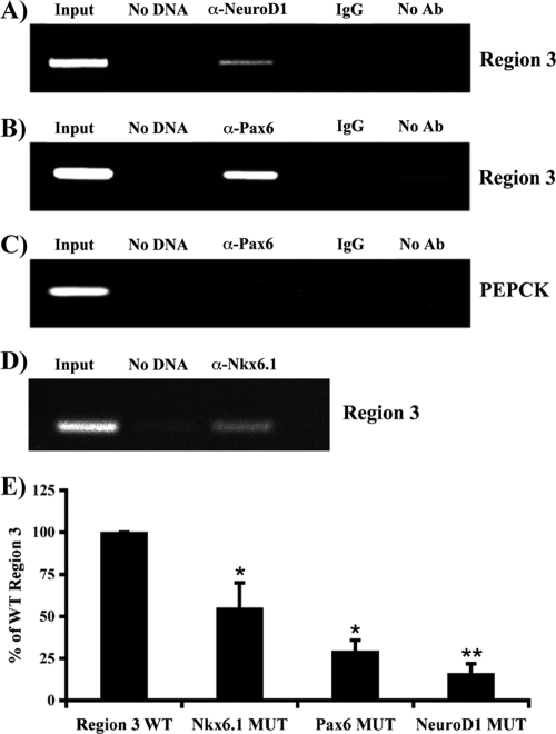
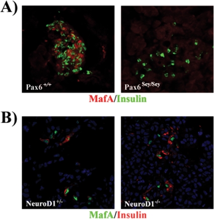
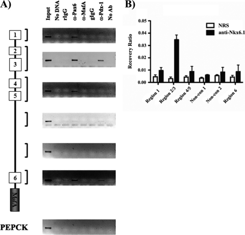
MafA is a key transcriptional activator of islet beta cells, and its exclusive expression within beta cells of the developing and adult pancreas is distinct among pancreatic regulators. Region 3 (base pairs -8118 to -7750 relative to the transcription start site), one of six conserved 5' cis domains of the MafA promoter, is capable of directing beta-cell-line-selective expression. Transgenic reporters of region 3 alone (R3), sequences spanning regions 1 to 6 (R1-6; base pairs -10428 to +230), and R1-6 lacking R3 (R1-6(DeltaR3)) were generated. Only the R1-6 transgene was active in MafA(+) insulin(+) cells during development and in adult cells. R1-6 also mediated glucose-induced MafA expression. Conversely, pancreatic expression was not observed with the R3 or R1-6(DeltaR3) line, although much of the nonpancreatic expression pattern was shared between the R1-6 and R1-6(DeltaR3) lines. Further support for the importance of R3 was also shown, as the islet regulators Nkx6.1 and Pax6, but not NeuroD1, activated MafA in gel shift, chromatin immunoprecipitation (ChIP), and transfection assays and in vivo mouse knockout models. Lastly, ChIP demonstrated that Pax6 and Pdx-1 also bound to R1 and R6, potentially functioning in pancreatic and nonpancreatic expression. These data highlight the nature of the cis- and trans-acting factors controlling the beta-cell-specific expression of MafA.


Figures

FIG. 1.
FIG. 1.
The MafA R1-6:eGFP and R1-6ΔR3:eGFP transgenes are expressed in similar tissue-specific manners. (A) Diagrams of the R3 (A), R1-6 (B), and R1-6ΔR3 (C) transgenic constructs. The start of transcription is from the hsp68 minimal heat shock promoter in R3:eGFP, while the endogenous start site in region 6 is utilized in the R1-6:eGFP and R1-6ΔR3:eGFP lines. Nuclear GFP was produced from all of the constructs (data not shown). (D) Whole-mount immunofluorescence of E13.5 embryos, performed with both R1-6:eGFP (E line is shown) and R1-6ΔR3:eGFP transgenic mouse lines. The expression patterns in these transgenic mouse lines were similar in most tissues, including the lens and limb buds (arrows), yet were absent in the spinal cord in R1-6ΔR3:eGFP mice (arrowhead). l, lens; d, digit tips; w, whisker hair shaft; h, hind limb; sc, spinal cord; BF, bright field; GFP, GFP fluorescence.
FIG. 2.
FIG. 2.
The MafA R1-6:eGFP transgene is active within the pancreas. Digestive organs, including the stomach, pancreas, spleen, and small intestine, were collected from the R1-6:eGFP A and E (shown) lines. The same developmental and adult GFP fluorescence patterns were obtained for both lines, with GFP not detected in the pancreatic buds until E15.5. The dorsal pancreatic bud is magnified (×4) in the E15.5 GFP inset. Pancreases are outlined in white. The punctate pattern in E15.5, E18.5, and P33 pancreases is characteristic of insulin+ cell expression. Low levels of EGFP were also detected in the E12.5 and E13.5 duodenum and stomach. BF, bright field; GFP, GFP fluorescence.
FIG. 3.
FIG. 3.
R3 is necessary for pancreatic transgenic expression. Gut tissues were collected from E13.5 and E18.5 embryos and P33 animals from the R-6ΔR3:eGFP line and examined via whole-mount immunofluorescence. No GFP expression was detected within the developing or adult pancreas, which is outlined in white. BF, bright field; GFP, GFP fluorescence.
FIG. 4.
FIG. 4.
MafA R1-6 drives GFP transgene expression, predominantly in MafA+ cells, in the developing and adult pancreas. Sections from E13.5 (A), E15.5 (B), E18.5 (C), and P33 (D) mice were collected, and sections were stained for GFP (green), MafA (red), and/or insulin (blue). GFP was first detected in MafA+ cells at E13.5. GFP was usually coexpressed with MafA (red arrowheads), although the occasional single GFP+ (white arrowheads) or MafA+ (yellow arrowheads) cell was observed, as illustrated in panel B. These images are representative of both the R1-6:eGFP A and E lines. Nuclei in panel A were counterstained in blue. Bar, 20 μm. (E) The mean percentages ± standard deviations of pancreatic GFP+ MafA+, GFP+, and MafA+ cells to the total number of GFP+ and MafA+ cells at E15.5 (red bars), E18.5 (blue bars), and P33 (gray bars) are shown for both the A and E lines. Cells were counted from at least three independently isolated pancreases per time point.
FIG. 5.
FIG. 5.
MafA transcription is induced in response to stimulating glucose treatment. Mouse islets were collected from the R1-6:eGFP line and cultured at a high (11 mM) or low (5 mM) glucose concentration. Real-time PCR was performed with primers specific to eGFP coding sequences, insulin II coding sequences, or the MafA 3′-untranslated region (UTR). Normalized mRNA values ± standard deviations are presented as fold changes between low and high glucose stimulation.
FIG. 6.
FIG. 6.
Identification of Pax6, NeuroD1, and Nkx6.1 binding sites within R3. (A) The gray highlighted sequences are identical between mouse and chicken R3 sequences. The locations of characterized and TRANSFAC-localized potential islet-enriched factor binding sites are superimposed on the R3 sequences. The Pdx-1, FoxA2, Nkx2.2, MafB, and Isl-1 binding sites (black boxes) have been described previously (2, 8, 27, 44). The Pax6, NeuroD1, and Nkx6.1 binding elements identified in the gel shift studies below are also denoted with black boxes. (B) Gel shifts were performed with probes corresponding to the consensus Pax6 site (lanes 1 and 2) and the four Pax6-like sites (lanes 3 to 14). The probes were incubated in the presence of βTC3 nuclear extract (alone; lanes 1, 3, 6, 9, and 12), and the specificity and composition of the βTC3 protein-DNA complexes were assayed with a 100-fold molar excess of the consensus Pax6 site (lanes 4, 7, 10, and 13) or by preincubation with an anti-Pax6 antibody (lanes 2, 5, 8, 11, and 14). (C) The βTC3 complexes formed with the NeuroD1-like site probe (bp −8046 to −8021; lane 1) were analyzed with a 10- to 50-fold molar excess of unlabeled wild-type competitor (R3 WT; lanes 2 to 4), a 25- to 50-fold molar excess of the R3 E-box mutant (R3 MT; lanes 5 and 6), or a 10-fold molar excess of the insulin NeuroD1 element (ICE WT; lane 7). Addition of anti-NeuroD1 antibody identified the specific location of the NeuroD1-containing binding complex (lane 8). (D) The Nkx6.1 consensus (lanes 5 to 8 and 13 to 15) and R3 Nkx6.1-like (lanes 1 to 4 and 9 to 12) sites were incubated in the presence of either βTC3 (lanes 1 to 8) or INS-1 (lanes 9 to 15) cell extracts. The Nkx6.1 complex was assessed with a 50-fold excess of either the wild-type R3 Nkx6.1 site (WT; lanes 2, 6, 10, and 14) or a probe for a R3 Nkx6.1 binding mutant (MUT; lanes 3, 7, and 11). The specificity of the Nkx6.1 complex was determined by incubation with an Nkx6.1 antibody (lanes 4, 8, 12, and 15). Asterisks denote nonspecific binding complexes in panels B to D.
FIG. 7.
FIG. 7.
Pax6, Nkx6.1, and NeuroD1 binding elements are necessary for R3 stimulation. Formaldehyde-cross-linked βTC3 chromatin was incubated with antibodies specific to NeuroD1 (A), Pax6 (B and C), and Nkx6.1 (D). Immunoprecipitated DNA was analyzed by PCR with primers specific to R3 (A, B, and D) or PEPCK (C). For controls, PCR was performed with total input DNA (1/100 dilution; input), no DNA, DNA immunoprecipitated with rabbit IgG (IgG), or DNA precipitated in the absence of antibody (no Ab). Only the anti-Pax6 results are shown for PEPCK, although the same pattern was seen for NeuroD1 and Nkx6.1. Each experiment was repeated with three separate chromatin preparations. (E) βTC3 cells were transfected with wild-type R3 (WT) and Pax6, NeuroD1, and Nkx6.1 binding-site mutants (MUT) of R3:pTk. Normalized region 3:pTk activity ± the standard error of the mean for each mutant is presented as a percentage of wild-type R3 activity. Wild-type and mutant R3:pTk constructs were active only in transfected β-cell lines, not in non-β cells (data not shown). Asterisks denote statistically significant differences between the mutant and wild-type activities, as assessed by Student's t test (*, P < 0.001; **, P < 0.005).
FIG. 8.
FIG. 8.
MafA is not produced in insulin+ cells remaining in Pax6 mutant mice but is present in those in NeuroD1 mutant mice. MafA and insulin expression was examined by immunofluorescence analysis of pancreatic sections from P2 Pax6sey/sey (A) and E15.5 NeuroD1−/− (B) mice and either wild-type or NeuroD1+/− control mice. NeuroD1+/− and wild-type mice were phenotypically indistinguishable (36), enabling us to use NeuroD1+/− mice as the control in this analysis. MafA was undetectable in the Pax6 mutant and was present in NeuroD1−/− insulin-producing cells. Nuclei were counterstained in blue.
FIG. 9.
FIG. 9.
Nkx6.1 binds only within R3, whereas Pdx-1 and Pax6 bind to multiple conserved regions of the MafA promoter. Scanning ChIP analysis was performed on roughly 10 kb of MafA, using antibodies specific to Pdx-1, MafA, and Pax6 (A) and to Nkx6.1 (B). (A) Immunoprecipitated DNA was analyzed with MafA-specific (R1, R2 and R3, R4 and R5, for distinct nonconserved sequences 1 and 2 and R6) and PEPCK (control)-specific primers. The PCRs were performed with total input DNA (1:100 dilution), no DNA, nonspecific rabbit IgG (rIgG), anti-Pax6, anti-MafA, nonspecific goat IgG (gIgG), anti-Pdx-1, or no antibody (no Ab). (B) Real-time PCR was performed on Nkx6.1-immunoprecipitated DNA. DNA samples were incubated with either Nkx6.1 or control IgG (NRS), with the data presented as recovery of the indicated region (region 1, regions 2 and 3, regions 4 and 5, or region 6), as a percentage of the input level. Each experiment was repeated with at least three independently isolated chromatin preparations.

Similar articles

Cited by

References

    1. Ahlgren, U., J. Jonsson, L. Jonsson, K. Simu, and H. Edlund. 1998. Beta-cell-specific inactivation of the mouse Ipf1/Pdx1 gene results in loss of the beta-cell phenotype and maturity onset diabetes. Genes Dev. 12:1763-1768. - PMC - PubMed
    1. Artner, I., B. Blanchi, J. C. Raum, M. Guo, T. Kaneko, S. Cordes, M. Sieweke, and R. Stein. 2007. MafB is required for islet beta cell maturation. Proc. Natl. Acad. Sci. U. S. A. 104:3853-3858. - PMC - PubMed
    1. Artner, I., Y. Hang, M. Marzur, T. Yamamoto, M. Guo, J. Linder, M. Magnuson, and R. Stein. MafA and MafB regulate genes critical to β cells in a unique temporal manner. Diabetes, in press. - PMC - PubMed
    1. Artner, I., J. Le Lay, Y. Hang, L. Elghazi, J. C. Schisler, E. Henderson, B. Sosa-Pineda, and R. Stein. 2006. MafB: an activator of the glucagon gene expressed in developing islet alpha- and beta-cells. Diabetes 55:297-304. - PubMed
    1. Ashery-Padan, R., X. Zhou, T. Marquardt, P. Herrera, L. Toube, A. Berry, and P. Gruss. 2004. Conditional inactivation of Pax6 in the pancreas causes early onset of diabetes. Dev. Biol. 269:479-488. - PubMed

Publication types

MeSH terms

LinkOut - more resources